This site uses cookies to improve your experience. To help us insure we adhere to various privacy regulations, please select your country/region of residence. If you do not select a country, we will assume you are from the United States. Select your Cookie Settings or view our Privacy Policy and Terms of Use.
Cookie Settings
Cookies and similar technologies are used on this website for proper function of the website, for tracking performance analytics and for marketing purposes. We and some of our third-party providers may use cookie data for various purposes. Please review the cookie settings below and choose your preference.
Used for the proper function of the website
Used for monitoring website traffic and interactions
Cookie Settings
Cookies and similar technologies are used on this website for proper function of the website, for tracking performance analytics and for marketing purposes. We and some of our third-party providers may use cookie data for various purposes. Please review the cookie settings below and choose your preference.
Strictly Necessary: Used for the proper function of the website
Performance/Analytics: Used for monitoring website traffic and interactions
IVC filters are used to prevent pulmonaryembolism in patients with venous thromboembolism and can’t receive anticoagulation treatment. An IVC filter is a small device that helps stop blood clots from going up into the lungs and is usually surgically inserted. years of insertion.
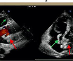
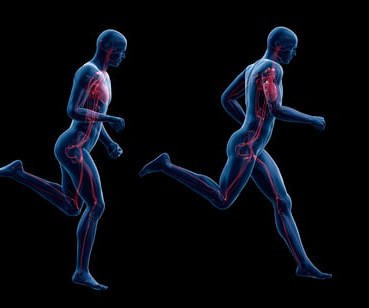
Notably, acute massive pulmonaryembolism (PE) with bilateral atrial thrombosis is an exceptional occurrence in CAPS. Acute pulmonaryembolism (PE) is a common cardiovascular disease that progresses rapidly and has a high mortality rate. It primarily affects small vessels, seldom impacting large vessels.
BACKGROUND:Prior clinical trials have demonstrated the efficacy of ultrasound-facilitated catheter-directed thrombolysis (USCDT) for the treatment of acute intermediate-risk pulmonaryembolism (PE) using reduced thrombolytic doses and shorter infusion durations.
Background D-Dimer testing is a diagnostic tool for exclusion of deep vein thrombosis (DVT) and pulmonaryembolism (PE). This study evaluated the diagnostic performance of the Tina-quant® D-Dimer Gen.2
Extended anticoagulant therapy with a reduced-dose of apixaban was noninferior to extended therapy with a full-dose of apixaban in preventing recurrent venous thromboembolism (VTE) in patients with active cancer and proximal deep-vein thrombosis or pulmonaryembolism, based on findings from the API-CAT trial presented at ACC.25
Genetic protein S (PS) deficiency caused by PROS1 gene mutation is an important risk factor for hereditary thrombophilia.Case introductionIn this case, we report a 28-year-old male patient who developed a severe pulmonaryembolism during his visit. Ultrasound showed no thrombosis in the veins of both lower limbs.
Phlegmasia cerulea dolens (PCD) is a rare yet severe complication of deep vein thrombosis (DVT), characterized by a high amputation rate and mortality. Early diagnosis and treatment are crucial in managing this condition. PCD predominantly affects the lower extremities rather than the upper extremities.
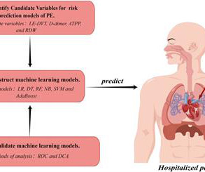
ResultsLogistic regression analysis identified lower extremity deep venous thrombosis, elevated D-dimer, shortened activated partial prothrombin time, and increased red blood cell distribution width as potential independent risk factors for PE. Clinical benefit was assessed using decision curve analysis (DCA).ResultsLogistic
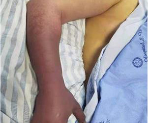
Inferior vena cava (IVC) agenesis is a rare congenital anomaly that has been implicated in up to 5% of unprovoked deep vein thrombosis (DVT) cases in young men under 30 years old. We present the case of a 28-year-old obese Caucasian male who arrived at our hospital with significant pain and swelling in his right lower extremity.
The Prevalence of Hypertension in Young Athletes: A Community-Based Screening Analysis Social Disparities, Experiences with Discrimination, and Cardiovascular Phenotypes in Black and White Collegiate American-Style Football Players Exercise-Induced Ventricular Fibrillation Cardiac Arrest in a Firefighter Using Intramuscular Testosterone with Segmental (..)
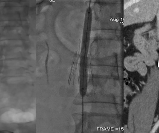
BackgroundThe VenaTech Convertible Vena Cava Filter (VTCF) is a device designed for insertion into the inferior vena cava (IVC) to prevent life-threatening pulmonaryembolism (PE).
A 42-year-old female with SLE, lupus cerebritis with related seizure disorder, and mesenteric venous thrombosis on warfarin initially presented for syncope. Given ongoing embolic phenomena, likely from LSE, she underwent MVR with mechanical valve and LA appendage ligation and continued mycophenolate and warfarin.
The CXR demonstrated no pulmonary edema. Sudden narrowing of a coronary artery due to ACS (plaque rupture with thrombosis and/or downstream showering of platelet-fibrin aggregates). This latter part has been implicated in embolic CVA. There was equally no anemia, sepsis, or hypoxia—only transient hypotension in the field.
In severe OHSS, increases in capillary permeability can result in hemoconcentration and hypercoagulability leading to thrombotic events, including stroke and cerebral venous thrombosis. Within the HCUP cohort, fewer than 10 patients (<1%) were hospitalized with a stroke or thrombotic event within 60 days of OHSS diagnosis.
Abstract: Venous thromboembolism (VTE), comprising deep-vein thrombosis (DVT) and pulmonaryembolism (PE), stands as the third leading cause of vascular-related mortality on a global scale.
to 10)), pulmonaryembolism (24.6 to 44.9)) and deep venous thrombosis (7.8 (4.3 Most studies had a high risk of bias. COVID-19 likely increases relative risk (RR (95% CI)) of myocardial infarction (3.3 (1.0 to 11.0)), stroke (3.5 (1.2 Other RTIs also likely increase the RR of myocardial infarction (2.9 (95% 95% CI 1.8
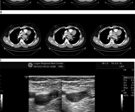
As in all ischemia interpretations with OMI findings, the findings can be due to type 1 AMI (example: acute coronary plaque rupture and thrombosis) or type 2 AMI (with or without fixed CAD, with severe regional supply/demand mismatch essentially equaling zero blood flow). CT angiogram showed extensive saddle pulmonaryembolism.
She had idiopathic ventricular fibrillation in 1992, treated with an EPD (Picture 1A), later replaced by a transvenous ICD.She was diagnosed with left femoral deep venous thrombosis and bilateral pulmonaryembolism and started on therapeutic anticoagulation. Despite empiric bronchial artery embolization, hemoptysis persisted.
Patients were drawn from neurology, cardiology, and other services. Descriptive statistics were used to compare trends across these groups.Results:Of the 3,966 patients, AF was the most common diagnosis (47.16% self-pay, 67.14% insured), followed by DVT and PE.
Adverse vascular outcomes used as endpoints include acute ischemic stroke, acute myocardial infarction, deep vein thrombosis/pulmonaryembolism, AF, and carotid artery dissection.A Patients with any adverse vascular outcomes before the index ECG were excluded. The mean age at the time of the index ECG was 44.3
In terms of complications, patients within the AKI cohort had lower rates of decompressive hemicraniectomy (1.37% vs. 2.38%, p = 0.52) and, interestingly, cerebral vasospasms (4.47% vs. 8.22%, p < 0.01).
CT angiogram chest: no aortic dissection or pulmonaryembolism. Serial chest xrays: progressive bilateral pulmonary edema. He spent several days in the PICU, undergoing workup including: Serial troponins: rising from 5,700 ng/L (unknown if I or T) to greater than 25,000 ng/L (greater than the lab's upper limit of reporting).
pulmonaryembolism, sepsis, etc.), Coronary thrombosis or embolism can result in MINOCA, either with or without a hypercoagulable state. Diagnosis of MINOCA should be made according to the Fourth Universal Definition of MI, in the absence of obstructive coronary artery disease (CAD) (no lesion ≥50%). myocarditis).
Women and black patients were less frequently treated with minimally invasive therapy compared to men or non-Black patients, according to new data from the REAL-PE analysis which investigated catheter-based pulmonaryembolism (PE) treatment. Late-breaking results from the study, for which Sahil A.
Introduction:COVID-19 infection has thus emerged to be a new risk factor for Cerebral Venous Thrombosis (CVT). Acute ischemic stroke (AIS), intracerebral hemorrhage (ICH), subarachnoid hemorrhage (SAH), epilepsy, deep vein thrombosis and pulmonaryembolism were secondary outcomes. vs 5.5%; HR=2.16; 95% CI=1.23-3.64
stroke), peripheral arterial disease, congenital heart anomalies, deep vein thrombosis, and pulmonaryembolism. Cardiovascular diseases (CVDs) encompass a range of disorders affecting the heart and blood vessels, such as coronary heart disease, cerebrovascular disease (e.g.,
However, after the procedure, moderate pericardial effusion developed in one patient (0.7%) and an acute pulmonaryembolism related to femoral vein thrombosis was observed in one patient (0.7%) during the first month. All of the patients had a >10 mm long-tunnel PFO.ResultsThe procedural success rate was 100%.
We organize all of the trending information in your field so you don't have to. Join thousands of users and stay up to date on the latest articles your peers are reading.
You know about us, now we want to get to know you!
Let's personalize your content
Let's get even more personalized
We recognize your account from another site in our network, please click 'Send Email' below to continue with verifying your account and setting a password.


Let's personalize your content