This site uses cookies to improve your experience. To help us insure we adhere to various privacy regulations, please select your country/region of residence. If you do not select a country, we will assume you are from the United States. Select your Cookie Settings or view our Privacy Policy and Terms of Use.
Cookie Settings
Cookies and similar technologies are used on this website for proper function of the website, for tracking performance analytics and for marketing purposes. We and some of our third-party providers may use cookie data for various purposes. Please review the cookie settings below and choose your preference.
Used for the proper function of the website
Used for monitoring website traffic and interactions
Cookie Settings
Cookies and similar technologies are used on this website for proper function of the website, for tracking performance analytics and for marketing purposes. We and some of our third-party providers may use cookie data for various purposes. Please review the cookie settings below and choose your preference.
Strictly Necessary: Used for the proper function of the website
Performance/Analytics: Used for monitoring website traffic and interactions
These are typical ECG changes that may indicate a pulmonaryembolism. The patient has an acute pulmonaryembolism. Sinus tachycardia may be present in acute pulmonaryembolism. ECG 2 was taken from the same patient 1 year earlier.
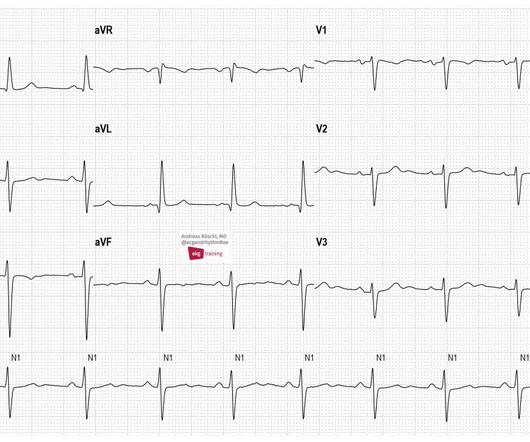
These are typical ECG changes that may indicate a pulmonaryembolism. The patient has an acute pulmonaryembolism. Sinus tachycardia may be present in acute pulmonaryembolism. Wee see a SR with LAFB and conspicuous T-wave inversions in the inferior leads and in V1-V6.
He was started on a heparin drip and CTA of the chest was ordered to rule out pulmonaryembolism. This is a case like many others posted (see list below) and the EKG from the patient’s original presentation can be quickly recognized as diagnostic for pulmonaryembolism. In fact, Kosuge et al. Accessed May 28, 2024.
male with pertinent past medical history including Atrial fibrillation, atrial flutter, cardiomyopathy, PulmonaryEmbolism, and hypertension presented to the Emergency Department via ambulance for respiratory distress and tachycardia. Bedside ultrasound showed volume depletion and no pulmonary edema. SVT with aberrancy?
Introduction Multiple abnormal electrocardiographic findings have been documented in patients experiencing acute pulmonaryembolism. Although sinus tachycardia is the most commonly encountered rhythmic disturbance, subsequent reports have highlighted other findings.
CT of the chest showed no pulmonaryembolism but bibasilar infiltrates. Even with tachycardia and a paced QRS duration of ~0.16 (And of course Ken's comments at the bottom) An elderly obese woman with cardiomyopathy, Left bundle branch block, and chronic hypercapnea presented hypoxic with altered mental status.
It shows sinus tachycardia with right bundle branch block. Taking a step back , remember that sinus tachycardia is less commonly seen in OMI (except in cases of impending cardiogenic shock). In patients with narrow QRS ( not this patient), this pattern is highly suggestive of acute pulmonaryembolism. Both were wrong.
Here is his ECG: Original image, suboptimal quality Quality improved with PM Cardio digitization The ECG is highly suggestive of acute right heart strain, with sinus tachycardia, S1Q3T3, and T wave inversions in anterior and inferior with morphology consistent with acute right heart strain. Moreover, there is tachycardia.
Smith interpretation: This is highly likely to be due to extreme right heart strain and is nearly diagnostic of pulmonaryembolism. It is of course pulmonaryembolism. No d-dimer or CT pulmonary angiogram was done when they discovered that she had normal coronary arteries. Junctional tachycardia? ).
Smith comment: before reading anything else, this case screamed pulmonaryembolism to me. I would do bedside ultrasound to look at the RV, look for B lines as a cause of hypoxia (which would support OMI, and argue against PE), and if any doubt persists, a rapid CT pulmonary angiogram. There is sinus tachycardia at ~100/minute.
Ken (below) is appropriately worried about pulmonaryembolism from the ECG. What I had not told him before he made that judgement is that the patient also had ultrasound B-lines of pulmonary edema. A Closer LOOK at the ECG in Figure-1: The ECG in Figure-1 shows sinus tachycardia — with QRS widening due to complete RBBB.
The morphology of V2-V4 is very specific in my experience for acute right heart strain (which has many potential etiologies, but none more common and important in EM than acute pulmonaryembolism). CT angiogram showed extensive saddle pulmonaryembolism. He had multiple cardiac arrests with ROSC regained each time.
The CXR demonstrated no pulmonary edema. An increase in myocardial oxygen demand due to tachycardia, elevated ventricular afterload (BP or Aortic stenosis), increased wall stretch, or a decrease in oxygen supply due to hypotension, anemia, hypoxia, sepsis, or a combination of all of the above. Type II ischemia.
Notice I did not say "pulmonaryembolism," because any form of severe acute right heart strain may produce this ECG. This includes, but is not limited to, PE, asthma/COPD exacerbation, hypoxic vasoconstriction from pneumonia, acute pulmonary hypertension exacerbation. The T-waves simply look different in Wellens'.
The ECG is rather classic for pulmonaryembolism, and indeed this was a large acute PE. This one is far more specific, as it is combined with sinus tachycardia and some T-wave inversion in V1-V3. and tachycardia, 1.8. this is highly suggestive of pulmonaryembolism. This is a classic S1Q3T3. Kosuge et al.
My answer: "This is classic for PE, but it can also be present in any hypoxia due pulmonary hypoxic vasoconstriction and resulting acute pulmonary hypertension and acute right heart strain. Tachycardia (or nearly) 2. An ECG was texted to me (Smith) without any clinical information: What did I say? This is NOT Wellens.
This usually represents posterior OMI, but in tachycardia and especially after cardiac arrest, this could simply be demand ischemia, residual subendocardial ischemia due to the low flow state of the cardiac arrest. This prompted cath lab activation. On arrival to the ED, this ECG was recorded: What do you think?
The bedside echo showed a large RV (Does this mean there is a pulmonaryembolism as the etiology?) When you suspect pulmonaryembolism due to large RV on POCUS, always look for right axis deviation and a large R-wave in V1 because the large RV may be entirely due to chronic RVH, not acute PE. Lots of info here.
Whenever you see tachycardia with bundle branch block, you should suspect that it is rate related BBB. First, as I have pointed out in posts on pulmonaryembolism (see links), T-wave inversion of anterior infarction (Wellens' syndrome) almost always has an upright T-wave in lead III. link] Shvilkin et al.
The patient had no hypertension, no tachycardia, a normal hemoglobin, no drug use, no hypotension/shock, no murmur of aortic stenosis. The patient had been on a long drive, suggesting possible pulmonaryembolism (this was unlikely given absence of tachyardia, hypoxia, or any other feature of PE), so we sent a d dimer.
No signs for aortic dissection or pulmonary embolus. --"Results were discussed with the ordering physician. If there are T-wave inversions and elevated trops in the context of persistent pain, think of other pathologies such as pulmonaryembolism. A CT Coronary angiogram was ordered. LAD plaque with 0-25 percent stenosis.
They include myocardial ischemia, acute pericarditis, pulmonaryembolism, external compression due to mass over the right ventricular outflow tract region, and metabolic disorders like hyper or hypokalemia and hypercalcemia. According to a recent systematic review and meta-analysis, spontaneous type 1 ECG had 2.4%
and the patient was converted to veno-venous (V-V) ECMO due to persistent pulmonary insufficiency. Clinically — despite an initial 2-fold increased troponin, the normal bedside Echo was reassuring against OMI or pulmonaryembolism. He remained supported on an intraaortic balloon pump. Here they are: Learning Points: 1.
Patients with pulmonaryembolism or aortic dissection who have normal variant ST elevation are at high risk of being diagnosed with pericarditis when what they have is far more serious!! The vast majority of cases with chest pain diffuse ST Elevation are due to Normal Variant ST Elevation, NOT to pericarditis.
If the patient has Abnormal Vital Signs (fever, hypotension, tachycardia, or tachypnea, or hypoxemia), then these are the primary issue to address, as there is ongoing pathology which must be identified. Serious outcomes included death, arrhythmia, myocardial infarction, structural heart disease, pulmonaryembolism, and hemorrhage.
Smith : This is classic for pulmonaryembolism (PE). Acute pulmonaryembolism was confirmed on CT angiogram: The patient did well. Cardiac Ultrasound may be a surprisingly easy way to help make the diagnosis Answer: pulmonaryembolism. Vitals were within normal limits. Now another, with ultrasound.
We organize all of the trending information in your field so you don't have to. Join thousands of users and stay up to date on the latest articles your peers are reading.
You know about us, now we want to get to know you!
Let's personalize your content
Let's get even more personalized
We recognize your account from another site in our network, please click 'Send Email' below to continue with verifying your account and setting a password.


Let's personalize your content