This site uses cookies to improve your experience. To help us insure we adhere to various privacy regulations, please select your country/region of residence. If you do not select a country, we will assume you are from the United States. Select your Cookie Settings or view our Privacy Policy and Terms of Use.
Cookie Settings
Cookies and similar technologies are used on this website for proper function of the website, for tracking performance analytics and for marketing purposes. We and some of our third-party providers may use cookie data for various purposes. Please review the cookie settings below and choose your preference.
Used for the proper function of the website
Used for monitoring website traffic and interactions
Cookie Settings
Cookies and similar technologies are used on this website for proper function of the website, for tracking performance analytics and for marketing purposes. We and some of our third-party providers may use cookie data for various purposes. Please review the cookie settings below and choose your preference.
Strictly Necessary: Used for the proper function of the website
Performance/Analytics: Used for monitoring website traffic and interactions
Discussion on pediatric exercise testing. Pediatric exercise testing may be used for evaluation of various disorders of cardiac rhythm rather than for inducible ischemia as in adults. In a child with suspected sinus node dysfunction, chronotropic incompetence from sinus node dysfunction can be assessed by exercise testing.
The primary end point was syncope recurrence, and the secondary end point was the reduction of the ventricular arrhythmia score during exercise testing. Twenty‐six PVT/ventricular fibrillation–triggering PVCs were identified for ablation. Induction of nontriggering PVCs after ablation is associated with a higher risk of syncope recurrence.
Abstract Introduction Fusion pacing requires correct timing of left ventricular pacing to right ventricular activation, although it is unclear whether this is maintained when atrioventricular (AV) conduction changes during exercise. QRSd during exercise ( p = .03), 03), peak O 2 pulse (mL/beat, a surrogate of stroke volume, p = .03),
LGE scar burden on MRI is thought to contribute to this risk, but its impact on electrophysiological substrate is not well understood. Hypertrophic cardiomyopathy (HCM) is associated with a predisposition to lethal ventricular arrhythmias.
Get Active, Stay Safe: Regular exercise is a cornerstone of heart health. Aim for at least 150 minutes of moderate-intensity exercise per week or 75 minutes of vigorous activity. However, during the summer, adjust your exercise routine to avoid the hottest part of the day. Know Your Limits: Listen to your body!
Get Active, Stay Safe: Regular exercise is a cornerstone of heart health. Aim for at least 150 minutes of moderate-intensity exercise per week or 75 minutes of vigorous activity. However, during the summer, adjust your exercise routine to avoid the hottest part of the day. Know Your Limits: Listen to your body!
Circulation: Arrhythmia and Electrophysiology, Ahead of Print. These include multiple environmental influences on QTc prolongation, exercise-induced repolarization abnormalities, and the profound implications of the constantly evolving nature of genetic testing and variant interpretation.
Electrophysiological study will show that, and this pathway can be ablated. Ebstein’s anomaly may present with a murmur for evaluation in the pediatric age group or in adults with arrhythmias or heart failure with cyanosis and exercise intolerance.
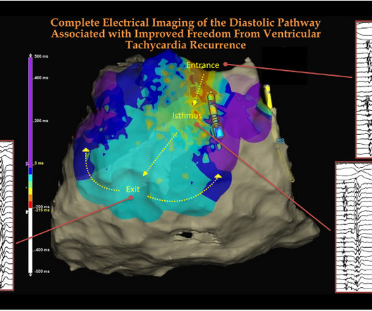
The term, VT mapping has been in vogue in clinical electrophysiology for more than half a century, right from Dr.Josephson and Wellens’ days. The learning point is that, in VT ablation, looking for anatomical diastolic tracts and its electrical activity becomes a key exercise. How can we remember this EP lesson easily ?
Abstract Introduction We report the case of a 37-year-old male athlete, who developed during exercise atrial and ventricular arrhythmias. No structural heart disease. Results Invasive programmed ventricular stimulation induced ventricular fibrillation. A heterozygous mutation in the CASQ2 gene (c.775G>T, 775G>T, p.E259X) was found.
Session 104) - What Is Really New in Electrophysiology That Will Change My Practice? The Guidelines Sessions at ACC.24 24: Joint American College of Cardiology/Journal of the American College of Cardiology Late-Breaking Clinical Trials (Session 402) Saturday, April 6 9:30 – 10:30 a.m.
It’s not just an isolated disorder of the heart’s rhythm, and we now know that the longer someone is in AFib, the harder it is to get them back to normal sinus rhythm,” said Jose Joglar, MD , professor of cardiac electrophysiology at UT Southwestern Medical Center in Dallas and chair of the writing committee.
Admission and referral to electrophysiology is always indicated. In this case, it was able to conduct at a rate of 257 (down the AV node, then up the bypass tract) 6. These tachydysrhythmias are so fast that they can degenerate into ventricular fibrillation. Unrecognized paroxysmal supraventricular tachycardia.
In addition to medication treatment, growing evidence is showing that the benefits of exercise outweigh the potential risks for patients with HCM. Low to moderate intensity recreational exercise should be part of how HCM patients manage their overall health.
This is mainly to account for the individual variation in anatomical location of right ventricular outflow tract, the main location of electrophysiological abnormalities in Brugada syndrome. Opinion is divided on the need for electrophysiology study.
Methods and Results A retrospective review of all treadmill exercise stress tests (TEST) was performed on patients with one of the three most common LQTS genotypes: LQT1, LQT2, and LQT3. This has not been demonstrated convincingly before because of the potentially confounding effects of beta blocker (BB) therapy.
5] The patient may experience pernicious exertional dyspnea, exercise intolerance, cough, or discrete fluid consolidation in the periphery – all red flag markers of clinical heart failure. Josephson’s Clinical Cardiac Electrophysiology: Techniques and Interpretations (6th ed). European Heart Journal, 28 , 2449-2455. [7] 7] Callans, D.
The present study aims to evaluate EKG changes during exercise stress tests in patients with BS and to identify any poor prognosis variables. No relationship was observed between the occurrence of arrhythmic events and ST-segment elevation during the exercise test. Methods This was an observational, case-control study.
ABSTRACT Background Exercise Oscillatory Ventilation (EOV) and a steep ventilatory efficiency (VE/VCO2) slope are features of ventilatory inefficiency on cardiopulmonary exercise testing (CPET), both associated with poor prognosis in patients with heart failure (HF). vs. 46.7mL/m 2 (39.8, 61.4), p =0.03].
We organize all of the trending information in your field so you don't have to. Join thousands of users and stay up to date on the latest articles your peers are reading.
You know about us, now we want to get to know you!
Let's personalize your content
Let's get even more personalized
We recognize your account from another site in our network, please click 'Send Email' below to continue with verifying your account and setting a password.


Let's personalize your content