This site uses cookies to improve your experience. To help us insure we adhere to various privacy regulations, please select your country/region of residence. If you do not select a country, we will assume you are from the United States. Select your Cookie Settings or view our Privacy Policy and Terms of Use.
Cookie Settings
Cookies and similar technologies are used on this website for proper function of the website, for tracking performance analytics and for marketing purposes. We and some of our third-party providers may use cookie data for various purposes. Please review the cookie settings below and choose your preference.
Used for the proper function of the website
Used for monitoring website traffic and interactions
Cookie Settings
Cookies and similar technologies are used on this website for proper function of the website, for tracking performance analytics and for marketing purposes. We and some of our third-party providers may use cookie data for various purposes. Please review the cookie settings below and choose your preference.
Strictly Necessary: Used for the proper function of the website
Performance/Analytics: Used for monitoring website traffic and interactions
Introduction Multiple abnormal electrocardiographic findings have been documented in patients experiencing acute pulmonary embolism. To date, only a limited number of cases involving a complete atrioventricular block have been reported in acute pulmonary embolism.
Some studies demonstrated that LVA ablation plus pulmonary veins isolation significantly improved the success rate. left atrial low-voltage areas (LVA) can significantly increase the risk of atrial fibrillation (AF) recurrence after catheter ablation. However, identification of LVA before the procedure is difficult.
A 77-year-old male with a history of two catheter ablation procedures, including pulmonary vein isolation and superior vena cava (SVC) isolation, presented with symptomatic palpitations. A twelve-lead electrocardiogram revealed atrial tachycardia (AT) with a cycle length of 240 ms.
This comprehensive evaluation included the use of ultrasound echocardiograms, computed tomography (CT) scans, electrocardiograms, mutagenesis analysis, and structural analysis to gain insights into the patient's condition and the underlying mechanisms of PD. Further genetic testing identified a homozygous mutation c.2662G>T
Objectives The aim of this study was to investigate echocardiographic parameters indicating reverse LA remodeling and potential associations with AF recurrence after pulmonary vein isolation (PVI). Electrocardiogram (ECG) test and transthoracic echocardiography were performed the day before and after PVI and again 3 months later.
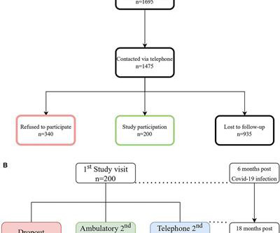
Follow-up approximately 6 months post discharge comprised a detailed patient history, clinical examination, transthoracic echocardiography, electrocardiogram, cardiac magnetic resonance imaging (cMRI), chest computed tomography (CT) scan, pulmonary function test (PFT), six-minute walk test (6MWT) and a laboratory panel.
In addition to pulmonary vein isolation (PVI), ablation of DISPERS was performed aiming at homogenizing, dissecting, isolating, or connecting DISPERS areas to nonconducting anatomical structures. Follow-up contained regular visits at our outpatient clinic at 1, 3, 6, and 12 months including 7-day Holter electrocardiograms. months).
Clinical evaluation and X-Ray chest showed features of pulmonary edema. Value of the electrocardiogram in localizing the occlusion site in the left anterior descending coronary artery in acute anterior myocardial infarction. ST segment elevation is noted in aVR. J Am Coll Cardiol. 2001 Nov 1;38(5):1348-54. Engelen DJ et al.
A deep neural network for 12-lead electrocardiogram interpretation outperforms a conventional algorithm, and its physician over-read, in the diagnosis of atrial fibrillation. The KEY to getting this patient better will doubtlessly include optimizing pulmonary function. IJC Heart and Vasculature 25(2019). Poon et al. sensitivity and 98.9%
You will note that it is essentially an unremarkable electrocardiogram except for some PACS. A majority of patients with MAT have longstanding pulmonary disease. Rather than antiarrhythmic medication — optimizing pulmonary function is the best treatment approach. The rhythm we see in the long lead II is not a common one.
In a study published in Communications Medicine , David Ouyang, MD, assistant professor of Cardiology and Medicine at Cedars-Sinai, along with Chugh and fellow investigators trained a deep learning algorithm to study patterns in electrocardiograms, also known as ECGs, which are recordings of the heart’s electrical activity.
Chronic Pulmonary Disease Lung diseases like chronic obstructive pulmonary disease (COPD) can lead to pulmonary hypertension, which in turn can cause the right side of the heart to enlarge, a condition known as cor pulmonale.
Among these, a fistula between the left anterior descending artery and the pulmonary artery is the rarest variant, comprising about 17% of all coronary artery fistula cases.Case:A 54-year-old male, with a known history of atrial fibrillation and hypertension, presented to our emergency department with non-rotatory dizziness.
Signify Research has just released a deep-dive qualitative analysis of developments around the use of AI to analyze and interpret electrocardiograms (ECGs), one of the world’s most ubiquitous diagnostic tests for cardiac disease.
CXR confirmed bilateral pulmonary edema and bilateral small effusions. 1211-1212 CrossRef View Record in Scopus Google Scholar 2 FI Marcus, W Zareba The electrocardiogram in right ventricular cardiomyopathy/dysplasia. I admitted her to cardiology with these concerns, and we agreed that cardiac MRI may help us confirm possible ARVC.
Santos Most Cited Article – Reduction in Hospitalization and Increase in Mortality Due to Cardiovascular Diseases during the COVID-19 Pandemic in Brazil Authors: Paulo Garcia Normando, José de Arimatéia Araujo-Filho, Gabriela de Alcântara Fonseca, Rodrigo Elton Ferreira Rodrigues, Victor Agripino Oliveira, Ludhmila Abrahão Hajjar, André Luiz (..)
Smith , d and Muzaffer Değertekin a DIFOCCULT: DIagnostic accuracy oF electrocardiogram for acute coronary OCClUsion resuLTing in myocardial infarction. On his physical examination, cardiac and pulmonary auscultation was completely normal. Bi-phasic scan showed no dissection or pulmonary embolism.
Emergency department Code STEMI patients with initial electrocardiogram labeled ‘normal’ by computer interpretation: a 7-year retrospective review. International evaluation of an artificial intelligence-powered electrocardiogram model detecting acute coronary occlusion myocardial infarction. J Electrocardiol 2017 2. Smith, Meyers.
Arrows indicate a higher number of elements/level in HFimpEF compared to the comparison population (pHFrEF). Events per 1000 person-years. # CV mortality is the composite endpoint for CV mortality + urgent transplant + left ventricular assist device implantation.
Explanation: Shown electrocardiogram suggests left ventricular hypertrophy. Shown electrocardiogram suggests left ventricular hypertrophy. Start aspirin and Plavix Correct answer: (B) (B) Echocardiogram is indicated. Hypertrophic cardiomyopathy is one of them. Start with a Free Trial.
At the bottom of the post, I have re-printed the section on aVR in my article on the ECG in ACS from the Canadian Journal of Cardiology: New Insights Into the Use of the 12-Lead Electrocardiogram for Diagnosing Acute Myocardial Infarction in the Emergency Department Case 1. Updates on the Electrocardiogram in Acute Coronary Syndromes.
Abnormal Electrocardiogram (ECG): Defined (San Fran syncope rule) as any new changes when compared to the last ECG or presence of non-sinus rhythm. Results : Electrocardiograms (99%), telemetry (95%), cardiac enzymes (95%), and head computed tomography (CT) (63%) were the most frequently obtained tests.
Transthoracic echocardiogram, bilateral carotid Doppler ultrasound, and electrocardiogram were normal. Cranial magnetic resonance imaging and magnetic resonance angiography showed no abnormalities. A treadmill exercise test revealed ischemic changes.
We organize all of the trending information in your field so you don't have to. Join thousands of users and stay up to date on the latest articles your peers are reading.
You know about us, now we want to get to know you!
Let's personalize your content
Let's get even more personalized
We recognize your account from another site in our network, please click 'Send Email' below to continue with verifying your account and setting a password.


Let's personalize your content