This site uses cookies to improve your experience. To help us insure we adhere to various privacy regulations, please select your country/region of residence. If you do not select a country, we will assume you are from the United States. Select your Cookie Settings or view our Privacy Policy and Terms of Use.
Cookie Settings
Cookies and similar technologies are used on this website for proper function of the website, for tracking performance analytics and for marketing purposes. We and some of our third-party providers may use cookie data for various purposes. Please review the cookie settings below and choose your preference.
Used for the proper function of the website
Used for monitoring website traffic and interactions
Cookie Settings
Cookies and similar technologies are used on this website for proper function of the website, for tracking performance analytics and for marketing purposes. We and some of our third-party providers may use cookie data for various purposes. Please review the cookie settings below and choose your preference.
Strictly Necessary: Used for the proper function of the website
Performance/Analytics: Used for monitoring website traffic and interactions
An electrocardiogram demonstrated sinus rhythm with T-wave alterations and a V2R/S ratio greater than 1. Despite this, the patient went on to develop chest pain, which was accompanied by electrocardiographic signs of acute extensive anterior wall myocardialinfarction and elevated troponin I levels.
Such a pattern is consistent with significant left main coronary artery stenosis. Angiography done after initial stabilization showed severe stenosis of distal left main coronary artery. ST segment elevation is noted in aVR. Clinical evaluation and X-Ray chest showed features of pulmonary edema. J Am Coll Cardiol. Engelen DJ et al.
The timely detection of myocardial fibrosis is crucial for intervention and improved outcomes. 68 Ga-FAPI-04 PET/CT shows promise in assessing fibroblast activation in patients with early myocardialinfarction characterized by prolonged myocardial ischemia. The results demonstrated tracer-specific uptake (SUVmax = 4.6)
Coronary computed tomography angiography (CTA) showed diffuse stenosis in the left anterior descending and the first diagonal branch arteries. Electrocardiogram (ECG) might not always show abnormalities, and chest pain is not always present. His headache improved after percutaneous coronary intervention.
Physician accuracy in interpreting potential ST-segment elevation myocardialinfarctionelectrocardiograms. He had 50% stenosis of the LAD which was deemed not culprit, and all other vessels were normal. I believe there is not quite enough STE for formal STEMI criteria, but some might measure 1.0 Carley et al.
Case A 43 year old male with a history of DM II, hyperlipidemia, and a family history of myocardialinfarction presented to a family clinic with two days of epigastric pain that started after consuming a meal. The attending provider wrote “Agree with electrocardiogram interpretation”. Normal EKG”. Normal ECG.
History sounds concerning for ACS (could be critical stenosis, triple vessel), but differential also includes dissection, GI bleed, etc. New insights into the use of the 12-lead electrocardiogram for diagnosing acute myocardialinfarction in the emergency department. His response: “subendocardial ischemia. Knotts et al.
As a brief review, HCM is a genetically inherited disorder that produces structural disarray in the myocardial cells. There is ventricular hypertrophy in the absence of abnormal loading conditions, such as aortic stenosis, or hypertension, for example – of which the most common variant is Asymmetric Septal Hypertrophy. 40; 1234-1241.
Signify Research has just released a deep-dive qualitative analysis of developments around the use of AI to analyze and interpret electrocardiograms (ECGs), one of the world’s most ubiquitous diagnostic tests for cardiac disease. Figure 1: The AI-ECG competitive vendor landscape New Horizons However, dramatic changes are afoot.
Diagnosis of Acute MyocardialInfarction in the Presence of Left Bundle Branch Block using the ST Elevation to S-Wave Ratio in a Modified Sgarbossa Rule. Electrocardiographic Diagnosis of Acute Coronary Occlusion MyocardialInfarction in Ventricular Paced Rhythm Using the Modified Sgarbossa Criteria.
At the bottom of the post, I have re-printed the section on aVR in my article on the ECG in ACS from the Canadian Journal of Cardiology: New Insights Into the Use of the 12-Lead Electrocardiogram for Diagnosing Acute MyocardialInfarction in the Emergency Department Case 1. Widimsky P et al. TIMI 0/1 flow).(61,62) Knotts et al.
Introduction:Cardio-cerebral infarction, a rare clinical presentation involving simultaneous acute ischemic stroke and acute myocardialinfarction, poses significant therapeutic challenges. The incidence of this dual infarction is currently unknown due to its rarity. ml subcutaneously once daily.
His father and brother both died of myocardialinfarction at ages 61 and 45, respectively. Advanced multi-vessel disease was found with stents deployed to the mid-LCx (80% stenosis), D1 (90% stensosis), and the pLAD (95% stenosis). A new electrocardiographic pattern indicating inferior myocardialinfarction.
Aortic Dissection, Valvular (especially Aortic Stenosis), Tamponade. Abnormal Electrocardiogram (ECG): Defined (San Fran syncope rule) as any new changes when compared to the last ECG or presence of non-sinus rhythm. heart auscultation (aortic stenosis); c. Abnormal ECG – looks for cardiac syncope. orthostatic vitals b.
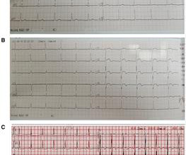
Wellens syndrome, characterised by specific T-wave changes on electrocardiogram (ECG), indicates critical proximal left anterior descending (LAD) stenosis and high acute myocardialinfarction risk. While revascularisation is the standard treatment, it may be unsuitable for elderly patients with comorbidities.
We organize all of the trending information in your field so you don't have to. Join thousands of users and stay up to date on the latest articles your peers are reading.
You know about us, now we want to get to know you!
Let's personalize your content
Let's get even more personalized
We recognize your account from another site in our network, please click 'Send Email' below to continue with verifying your account and setting a password.


Let's personalize your content