This site uses cookies to improve your experience. To help us insure we adhere to various privacy regulations, please select your country/region of residence. If you do not select a country, we will assume you are from the United States. Select your Cookie Settings or view our Privacy Policy and Terms of Use.
Cookie Settings
Cookies and similar technologies are used on this website for proper function of the website, for tracking performance analytics and for marketing purposes. We and some of our third-party providers may use cookie data for various purposes. Please review the cookie settings below and choose your preference.
Used for the proper function of the website
Used for monitoring website traffic and interactions
Cookie Settings
Cookies and similar technologies are used on this website for proper function of the website, for tracking performance analytics and for marketing purposes. We and some of our third-party providers may use cookie data for various purposes. Please review the cookie settings below and choose your preference.
Strictly Necessary: Used for the proper function of the website
Performance/Analytics: Used for monitoring website traffic and interactions
Myocardial ischemia may induce myocardial fibrosis, a condition that progressively leads to ventricular remodeling, heightening the risk of heart failure. 68 Ga-FAPI-04 PET/CT shows promise in assessing fibroblast activation in patients with early myocardial infarction characterized by prolonged myocardial ischemia.
The Kardia 12L ECG System, featuring a game-changing patented technology, is the world’s first AI-powered handheld 12-lead electrocardiogram ( ECG ) system with a unique single-cable design. This is the world’s first AI that can detect life-threatening cardiac conditions, including heart attacks, using a reduced leadset.
Publication date: 15 March 2024 Source: The American Journal of Cardiology, Volume 215 Author(s): Claudia Algaze, Henry Chubb, Anna M. Deitch, Thomas Collins
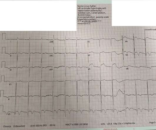
The ECG did not meet STEMI criteria, and the final cardiology interpretation was “ST and T wave abnormality, consider anterior ischemia”. Hence the first ECG was labeled 'anterior ischemia' based on ST depression, rather than identifying this as reciprocal from posterior OMI. But are there any other signs of Occlusion MI? Meyers et al.
4,5] We have now formally studied this question: Emergency department Code STEMI patients with initial electrocardiogram labeled ‘normal’ by computer interpretation: a 7-year retrospective review.[6] Other signs of OMI that complement the ECG include new regional wall motion abnormalities and refractory ischemia References 1.
STE limited to aVR is due to diffuse subendocardial ischemia, but what of STE in both aVR and V1? The additional ST Elevation in V1 is not usually seen with diffuse subendocardial ischemia, and suggests that something else, like STEMI from LAD occlusion, could be present. Was this: 1) ACS with ischemia and spontaneous reperfusion?
Precordial ST depression may be subendocardial ischemia or posterior STEMI. I have warned in the past that one must think of other etiologies of ischemia when there is tachycardia. Whether it is subendocardial ischemia or posterior STEMI, if you cannot get it to resolve, you must activate the cath lab. There is no ST elevation.
Chest Pain Severity Rating Is a Poor Predictive Tool in the Diagnosis of ST-Segment Elevation Myocardial Infarction [link] Abstract Current ST-segment elevation myocardial infarction (STEMI) guidelines require persistent electrocardiogram ST-segment elevation, cardiac enzyme changes, and symptoms of myocardial ischemia.
Mechanism is thought to be due to sustained sympathetic stimulation, probably caused by dysfunction of insular cortex resulting in reversible neurogenic damage to the myocardium which could include contraction bands and subendocardial ischemia [2]. Lead electrocardiogram changes after supratentorial intracerebral hemorrhage.
This may result in ischemia (lack of oxygen to the heart muscle), causing parts of the heart to weaken and enlarge. Electrocardiogram (ECG/EKG) An ECG records the electrical activity of the heart and can help detect abnormalities in the heart’s rhythm that might contribute to enlargement.
His response: “subendocardial ischemia. Smith : It should be noted that, in subendocardial ischemia, in contrast to OMI, absence of wall motion abnormality is common. With the history of Afib, CTA abdomen was ordered to r/o mesenteric ischemia vs ischemic colitis vs small bowel obstruction. Anything more on history?
There is appreciable STE aVR with near-global STD that appropriately maximizes in Leads II and V5, and thus suggesting a circumstance of generic, diffusely populated, circumferential subendocardial ischemia versus occlusive coronary thrombus. [1] There is evolution from Wellens Pattern A to Pattern B, now inclusive of V6.
The patient with no prior cardiac history presented in the middle of the night with acute chest pain, and had this ECG recorded during active pain: I did not see any ischemia on this electrocardiogram. It does not look entirely normal, since there are some nonspecific STT abnormalities, such as flattening of the T waves in aVL.
Post by Smith and Meyers Sam Ghali ( [link] ) just asked me (Smith): "Steve, do left main coronary artery *occlusions* (actual ones with transmural ischemia) have ST Depression or ST Elevation in aVR?" That said, complete LM occlusion would be expected to have subepicardial ischemia (STE) in these myocardial territories: STE vector 1.
An initial electrocardiogram (ECG) is provided below. Learning Point: Concordant ST segment elevation can arise from profound ischemia triggered by ventricular tachycardia (VT), or it may represent an exaggerated basal ST change accompanying tachycardia. The patient was promptly admitted to the hospital for further evaluation.
5] Back to the case The patient had serial ECGs over the next hour with no significant change: The first troponin came back at 1,400 ng/L (normal <26 in males and <16 in females), confirming MI – and the patient’s refractory ischemia indicated this was an Occlusion MI. Clin Cardiol 2022 4. Herman, Meyers, Smith et al.
Pediatric exercise testing may be used for evaluation of various disorders of cardiac rhythm rather than for inducible ischemia as in adults. Discussion on pediatric exercise testing. In a child with suspected sinus node dysfunction, chronotropic incompetence from sinus node dysfunction can be assessed by exercise testing.
An electrocardiogram is a machine used to record the heart's electrical activity. Electrocardiogram, echocardiogram, and some other tests are done for patients with cardiac arrest. Poor blood supply Ischemia, or inadequate blood supply to the heart, is an abnormality that can be detected in an ECG test.
The ECG in the chart was read as "no obvious ST changes," (even though no previous ECG was available) and the formal read by the emergency physicians was: "ST deviation and moderated T-wave abnormality, consider lateral ischemia." When the ischemia is resolved, the wall motion may completely recover, or there may be persistent stunning.
Computer read: "Non-specific ST abnormality, consider anterior subendocardial ischemia" There are very poor R-waves in V1-V4 suggesting old anterior MI. Firstly, subendocardial ischemia does not localize on 12-Lead ECG. But the real question at hand is: Are these precordial ST-depressions a result of subendocardial ischemia?
There is broad subendocardial ischemia as demonstrated by STE aVR with concomitant STD that almost appears appropriately maximal in Leads II and V5. There is LBBB-like morphology with persistent patterns of subendocardial ischemia. This is the initial ECG: The QRS is widened with a regular cadence, and there are no discernable P waves.
ST segment elevation in aVR in proximal LAD occlusion before first septal is thought to be due to transmural ischemia of the basal part of the septum. Value of the electrocardiogram in localizing the occlusion site in the left anterior descending coronary artery in acute anterior myocardial infarction. J Am Coll Cardiol.
This was just published in JAMA Internal Medicine: The de Winter Electrocardiogram Pattern Evolving From Hyperacute T Waves It reminded me that many believe, due to the assertions in the original de Winter's article, that de Winter's waves are stable.
When “spot diagnosing” precordial ST-depression I instinctively evaluate aVR for any corresponding ST-elevation to see if an emerging pattern of broad subendocardial ischemia can be appreciated, in which the ST-depression should be otherwise global and demonstrably maximal in Leads II and V5. ST-elevation, etc.) is present. 5] Meyers, H.
She requires maximal medical management per all current guidelines (including heparin and P2Y12 inhibitor per cardiology), as well as consideration for emergent cath in the case of persistent ischemia. So what will you do for this patient? They found an acute, total, thrombotic occlusion of the proximal LAD. They opened it. Patel et al.,
EKG shown here: LAFB with no clear signs of OMI or ischemia. Interestingly, this patient was seen in the ED for hypertension and headache 3 days earlier. No labs were performed. EKG and CT head were performed. Imaging was negative and he was discharged home.
In other words, the inferior ST segments in the first ECG show more straightening which is more concerning for ischemia. Below is the post -PCI electrocardiogram. Most notably the ST depression in the inferior leads is slightly more upsloping. The QoH interpretation however was the same for both ECGs.
The stress electrocardiogram is non-diagnostic. This ST-T wave appearance in the lateral chest leads of ECG #2 is consistent with L V “ S train” vs ischemia. No wall motion abnormality at rest. No wall motion abnormality with stress. Next day, a stress echo was done: The exercise stress echocardiogram is normal.
The patient continued to have ischemia after PCI, and in fact had an episode of polymorphic VT shortly after while in the ICU. Clinical value of 12-lead electrocardiogram after successful reperfusion therapy for acute myocardial infarction. This was recorded 2.5 Reexamining the "gold standard" for myocardial reperfusion treatment.
Background: The value of the 12-lead ECG in the diagnosis of non-ST-elevation myocardial infarction (NSTEMI) is limited due to insufficient sensitivity and specificity of standard markers of ischemia and because ECG confounders may prevent their application.
More likely, the patient had crescendo angina, with REVERSIBLE ischemia for 48 hours that only became potentially irreversible (STEMI) at that point in time. During the 48 hours of angina, such reversible ischemia often leads to myocardial stunning with akinesis of the myocardial wall that puts it at risk for thrombus.
Electromechanical association: a subtle electrocardiogram artifact. Arterial pulse tapping artifact [link] This online article references the article below by Emre Aslanger, a great guy who occasionally corresponds with me about ECGs. Aslanger E, Yalin K. Journal of Electrocardiology. 2012;45(1):15-17. doi:10.1016/j.jelectrocard.2010.12.162.
These findings are concerning for inferior wall ischemia with possible posterior wall involvement. You will note that it is essentially an unremarkable electrocardiogram except for some PACS. The morphology in V2 is also concerning and it appears that the ST segment is being pushed down, as in ST depression.
A Deep Neural Network learning algorithm outperforms a conventional algorithm for emergency department electrocardiogram interpretation. This ECG comes from Pierre Taboulet ( [link] /)( [link] ) an ECG whiz who codes a lot of ECGs for Cardiologs' Artificial Intelligence Deep Neural Network algorithm ( [link] ). What an honor.
Across both selected patient populations, the positive predictive value was highest in patients with chest pain, with ischaemia on the electrocardiogram, and with a history of ischaemic heart disease. to 62.2%) in the UK and 4.2% (68/1631) and 16.4% (13.0% to 20.3%) in the US.
Long-term Follow-up of Patients with Brugada Syndrome from a Tertiary Referral Center in Iran Abstract Background Brugada syndrome (BrS) is characterized by ST-segment elevation in the right precordial leads, which is not explained by ischemia, electrolyte disturbances, or obvious structural heart disease. 01), longer PR interval ( p =.03),
Integrating ECG Testing to identify cardiometabolic changes and measure the risk of CVD Electrocardiogram Machines like , Wellnest 12L Pro2 serve as a valuable tools in this endeavor, offering a window into the intricate relationship between ART and cardiovascular health. ECGs can identify these abnormalities, enabling timely intervention.
Abnormal Electrocardiogram (ECG): Defined (San Fran syncope rule) as any new changes when compared to the last ECG or presence of non-sinus rhythm. Evidence of acute ischemia (may be subtle) vii. Electrocardiogram-based risk stratification was useful in guiding the use of specialized cardiovascular tests. __ 9) François P.
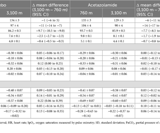
Exercise electrocardiograms were recorded at the National Center of Internal Medicine and Cardiology, Bishkek (760m) and on the day of arrival at the Tuja Ashu high-altitude clinic (3,100m), Kyrgyzstan. The mean difference (95% CI) in STE between post-peak exercise between 3,100m and 760m was 0.22mm (0.06 to 0.39) and 0.09mm (0.06
BackgroundPainful left bundle branch block (LBBB) syndrome is an uncommon disease that is defined as intermittent episodes of angina associated with simultaneous LBBB changes on an electrocardiogram (ECG) with the absence of flow-limiting coronary artery disease or ischemia on functional testing.
Artificial intelligence (AI) algorithms show promise to improve electrocardiogram (ECG) interpretation. Methods Electrocardiograms were categorized by (1) STEMI criteria, (2) ECG integrated device software and (3) a proprietary AI algorithm (Queen of Hearts (QOH), Powerful Medical).
The de Winter electrocardiogram pattern is an infrequent presentation, reported to occur in 2% to 3.4% In this situation, even after the ischemia is relieved and myocardial blood flow is restored myocardial contractile function remains impaired for a variable period of time (usually days to a few weeks). References: Kloner, R.
We organize all of the trending information in your field so you don't have to. Join thousands of users and stay up to date on the latest articles your peers are reading.
You know about us, now we want to get to know you!
Let's personalize your content
Let's get even more personalized
We recognize your account from another site in our network, please click 'Send Email' below to continue with verifying your account and setting a password.


Let's personalize your content