This site uses cookies to improve your experience. To help us insure we adhere to various privacy regulations, please select your country/region of residence. If you do not select a country, we will assume you are from the United States. Select your Cookie Settings or view our Privacy Policy and Terms of Use.
Cookie Settings
Cookies and similar technologies are used on this website for proper function of the website, for tracking performance analytics and for marketing purposes. We and some of our third-party providers may use cookie data for various purposes. Please review the cookie settings below and choose your preference.
Used for the proper function of the website
Used for monitoring website traffic and interactions
Cookie Settings
Cookies and similar technologies are used on this website for proper function of the website, for tracking performance analytics and for marketing purposes. We and some of our third-party providers may use cookie data for various purposes. Please review the cookie settings below and choose your preference.
Strictly Necessary: Used for the proper function of the website
Performance/Analytics: Used for monitoring website traffic and interactions
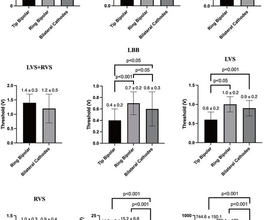
The present study aimed to compare the electrophysiological characteristics of LBBP in different bilateral electrode pacing vector configurations.MethodsA total of 57 patients who met the criteria for left bundle branch (LBB) capture and underwent three bilateral electrode pacing vector configuration test were enrolled.
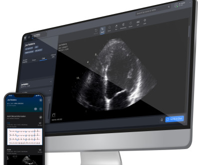
In a study published in Communications Medicine , David Ouyang, MD, assistant professor of Cardiology and Medicine at Cedars-Sinai, along with Chugh and fellow investigators trained a deep learning algorithm to study patterns in electrocardiograms, also known as ECGs, which are recordings of the heart’s electrical activity.
Notwithstanding many insightful observations, the electrocardiogram (ECG) arguably ignited the big bang in our understanding of cardiac arrhythmias. The development of electrophysiology (EP) studies through intravascular catheters provided the leap to verifying mechanisms, setting the stage for interventions.
Viz HCM uses artificial intelligence to analyze all 12-lead electrocardiograms (ECGs) from across a health system to identify suspected HCM cases, notify cardiology care teams and increase the likelihood that patients get the right follow-up and diagnosis. JACC: Clinical Electrophysiology.
The interrupted technique of left bundle branch pacing (LBBP) limits the continuous monitoring of paced electrocardiogram (ECG) and intracardiac electrogram (EGM) transitions, which may result in overlooked or misinterpreted subtle transitions.
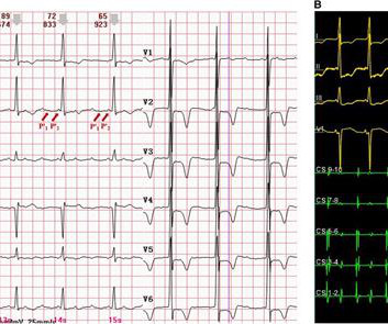
However, widely split P' waves in focal atrial tachycardia (AT) on a surface electrocardiogram (ECG) have rarely been reported. The electrophysiological mechanism is relatively difficult to clarify, requiring a electrophysiological study. Background Widely split P waves in sinus rhythm have been reported previously.
Rapid changes in both device technology and the field of electrophysiology (EP) necessitate a growing need for continuing medical education and intraoperative support.
He was not on any medication and his baseline electrocardiogram (ECG) was normal. During the electrophysiologic study, diagnostic catheters were placed in coronary sinus (CS), His bundle - right bundle (HB-RB) region and right ventricular apex (RVA).
Methods and Results A 21-year-old female with supraventricular tachycardia (SVT) and pre-excitation on electrocardiogram (ECG) underwent electrophysiology study (EPS) confirming an AS-AP with anterograde and retrograde conduction. Retrograde conduction was still present and confirmed by repeating electrophysiological maneuvers.
It is also published in Heart Rhythm , the official journal of the HRS, Journal of Arrhythmia , the official journal of the APHRS, and Journal of Interventional Cardiac Electrophysiology , the official journal of the LAHRS. 7 Atrial fibrillation has a significant impact on people’s lives.
A 12-lead electrocardiogram revealed a narrow QRS complex tachycardia with a rate of 157 beats per minute and a prolonged RP relationship. Subsequent electrophysiological study findings identified the tachycardia as originating from the anterior limbus of the PFO. Echocardiography indicated a severely reduced ejection fraction of 22%.
Circulation: Arrhythmia and Electrophysiology, Ahead of Print. BACKGROUND:It is difficult to identify patients with atrial fibrillation (AF) most likely to respond to ablation. We compared 6 machine learning models to predict acute and long-term end points after ablation and used Shapley explainability analysis to contrast phenotypes.
We report the case of a 66-year-old man affected by metastatic lung adenocarcinoma with type 1 Brugada phenocopy at electrocardiogram (ECG) after initiation of an oncological therapy with BRAF and MEK inhibitors. ABSTRACT Management of patients with drug-induced type 1 Brugada pattern is complex and controversial.
This measurement has been correlated with those made at electrophysiology study and may predict the potential risk of rapid anterograde conduction if the person develops atrial fibrillation. Response of accessory pathway conduction to exercise gives useful information on the rate at which anterograde preexcitation is blocked.
Signify Research has just released a deep-dive qualitative analysis of developments around the use of AI to analyze and interpret electrocardiograms (ECGs), one of the world’s most ubiquitous diagnostic tests for cardiac disease.
Advanced arrhythmic substrate consisting of significant conduction abnormalities due to inflammation and fibrosis can be identified by specific electrocardiogram signs, such as fragmented QRS and ε wave. Methods The study population included consecutive 52 patients with CS and sustained VTA. Twenty-five out of 52 patients experienced ES.
The demographic, clinical, and electrocardiogram features of the patients in each group were compared. For symptomatic patients, the correlation between palpitations and PVC was further evaluated based on the temporal consistency of symptom onset and PVC occurrence. Results Of the 214 patients enrolled, 124(57.9%) experienced palpitations.
Preimplant electrocardiogram screening is recommended to prevent implantation in patients at high risk of T wave over-sensing. The S-ICD is implanted subcutaneously or intramuscularly with the generator placed in the left midaxillary line and the lead tunneled subcutaneously in the left para-sternal region.
Electromechanical association: a subtle electrocardiogram artifact. Acute chest pain and a bizarre ECG Bizarre (Hyperacute??) T-waves More info on arterial pulse tapping artifact [link] Aslanger E, Yalin K. Journal of Electrocardiology. 2012;45(1):15-17. doi:10.1016/j.jelectrocard.2010.12.162. 2010.12.162. 17, 2023 post ).
The 12-lead electrocardiogram (ECG) and three-dimensional (3D) electroanatomical maps were analyzed. Methods This study enrolled 10 patients who underwent radiofrequency catheter ablation for a typical AFL. Electroanatomical mapping was performed both during typical AFL and entrainment from the right atrial appendage (RAA).
FUJI trial, we assessed the differences in electrocardiogram (ECG) parameters during RV pacing between a delivery catheter system and a stylet system and their associations with the lead tip positions. In this subanalysis of the Mt.
Follow-up contained regular visits at our outpatient clinic at 1, 3, 6, and 12 months including 7-day Holter electrocardiograms. In addition to pulmonary vein isolation (PVI), ablation of DISPERS was performed aiming at homogenizing, dissecting, isolating, or connecting DISPERS areas to nonconducting anatomical structures. to 202.2 ± 21.6 ms
She has not yet been seen by electrophysiology or had further genetic testing for Brugada syndrome. Induced Brugada-type electrocardiogram, a sign for imminent malignant arrhythmias. As for our patient, on discharge, her EKG had completed returned to her baseline morphology and she has been doing well in follow-up.
She has not yet been seen by electrophysiology or had further genetic testing for Brugada syndrome. Induced Brugada-type electrocardiogram, a sign for imminent malignant arrhythmias. As for our patient, on discharge, her EKG had completed returned to her baseline morphology and she has been doing well in follow-up.
Abnormal Electrocardiogram (ECG): Defined (San Fran syncope rule) as any new changes when compared to the last ECG or presence of non-sinus rhythm. Results : Electrocardiograms (99%), telemetry (95%), cardiac enzymes (95%), and head computed tomography (CT) (63%) were the most frequently obtained tests.
Thirty-three (43%) patients had a typical spontaneous electrocardiogram (ECG) pattern. Results Of the 76 cases, 79% were proband and 21% were detected during screening after diagnosis of BrS in a family member. 01), longer PR interval ( p =.03), 03), and SCN5A mutation ( p = .01) 01) than asymptomatic patients.
A score including ECG pattern, early familial SCD antecedents, inducible electrophysiological study, presentation as syncope or as aborted SCD and SND had a predictive performance of 0.82. and proband status (HR 2.1). A score greater than 2 conferred a 5-year event probability of 9.2%.
We organize all of the trending information in your field so you don't have to. Join thousands of users and stay up to date on the latest articles your peers are reading.
You know about us, now we want to get to know you!
Let's personalize your content
Let's get even more personalized
We recognize your account from another site in our network, please click 'Send Email' below to continue with verifying your account and setting a password.


Let's personalize your content