This site uses cookies to improve your experience. To help us insure we adhere to various privacy regulations, please select your country/region of residence. If you do not select a country, we will assume you are from the United States. Select your Cookie Settings or view our Privacy Policy and Terms of Use.
Cookie Settings
Cookies and similar technologies are used on this website for proper function of the website, for tracking performance analytics and for marketing purposes. We and some of our third-party providers may use cookie data for various purposes. Please review the cookie settings below and choose your preference.
Used for the proper function of the website
Used for monitoring website traffic and interactions
Cookie Settings
Cookies and similar technologies are used on this website for proper function of the website, for tracking performance analytics and for marketing purposes. We and some of our third-party providers may use cookie data for various purposes. Please review the cookie settings below and choose your preference.
Strictly Necessary: Used for the proper function of the website
Performance/Analytics: Used for monitoring website traffic and interactions
Electrocardiogram results showed sinus tachycardia, QRS widening, low-voltage complexes, and ST-segment elevation. A woman in her mid-20s presented with acute fever, chest pain, and exertional dyspnea. What would you do next?
In a study published in Communications Medicine , David Ouyang, MD, assistant professor of Cardiology and Medicine at Cedars-Sinai, along with Chugh and fellow investigators trained a deep learning algorithm to study patterns in electrocardiograms, also known as ECGs, which are recordings of the heart’s electrical activity.
Chen Yan and researcher Sun Qibin from the University of Science and Technology of China (USTC) achieved contactless electrocardiogram (ECG) monitoring through a millimeter-wave radar system. Recently, a team led by Prof. Their work was published in IEEE Transactions on Mobile Computing and reported by IEEE Spectrum.
Artificial intelligence (AI)-enabled sinus rhythm (SR) electrocardiogram (ECG) interpretation can aid in identifying undiagnosed paroxysmal atrial fibrillation (AF) in patients with embolic stroke of undetermined source (ESUS).
This results in severe chest pain or discomfort, with the subsequent release of cardiac biomarkers, and alterations in the electrocardiogram. It can cause diminished heart function and mortality if not treated properly with suitable measures.
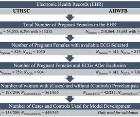
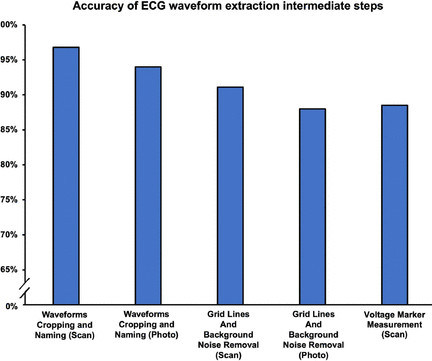
In this study, we developed artificial intelligence models to detect and predict preeclampsia from electrocardiograms (ECGs) in point-of-care settings. Early diagnosis and management of preeclampsia can improve outcomes for both mother and baby.
To diagnose heart conditions including heart attacks and heart rhythm disturbances, clinicians typically rely on 12-lead electrocardiograms (ECGs) -- complex arrangements of electrodes and wires placed around the chest and limbs to detect the heart's electrical activity.
Background Researchers have developed machine learning-based ECG diagnostic algorithms that match or even surpass cardiologist level of performance. However, most of them cannot be used in real-world, as older generation ECG machines do not permit installation of new algorithms.
That's why the recent and rapid rise in wearable electronic health-monitoring devices with heart rate-measuring electrocardiograms (ECG) represents a significant step forward. Nearly 200 million people around the globe have coronary heart disease, which accounts for about one in every six deaths, according to the British Heart Foundation.
Current clinical methods to identify the SOO are based on qualitative analysis of pre-operative electrocardiograms (ECG), heavily relying on physician’s expertise. Pinpointing the SOO enhances the likelihood of a successful procedure, reducing intervention times and recurrence rates.
Electrocardiogram tests may someday be used with an artificial intelligence (AI) model to detect premature aging and cognitive decline, according to a preliminary study presented at the American Stroke Association's International Stroke Conference 2025, held in Los Angeles, Feb. 57, 2025.
Investigators trained an AI model on pre-operative electrocardiograms, discovering a new use for the 130-year-old test. Invented in the late-1800s, an electrocardiogram is a commonly deployed test that involves placing electrodes on the skin to measure the heart’s electrical activity and assess how well the heart is functioning.
Biological age can be predicted using artificial intelligence (AI) trained on electrocardiograms (ECGs), which is prognostic for mortality and cardiovascular events.
If zero-shot VQA can be applied to a 12-lead electrocardiogram (ECG), a prevalent diagnostic tool in the medical field, the potential benefits to the field would be substantial. Large Language Models (LLM) are increasingly multimodal, and Zero-Shot Visual Question Answering (VQA) shows promise for image interpretation.
electrocardiogram, electroencephalogram, cardiac monitor), creating artifacts that hinder their interpretation. In emergencies where obtaining an electrocardiogram is crucial, the need for more consensus on reducing electrical artifacts in patients with DBS becomes a significant challenge.
Single-lead electrocardiograms (1L ECG) are increasingly used for atrial fibrillation (AF) detection. Automated 1L ECG interpretation may possess prognostic value for future AF among cases where screening does not result in a short-term AF diagnosis.
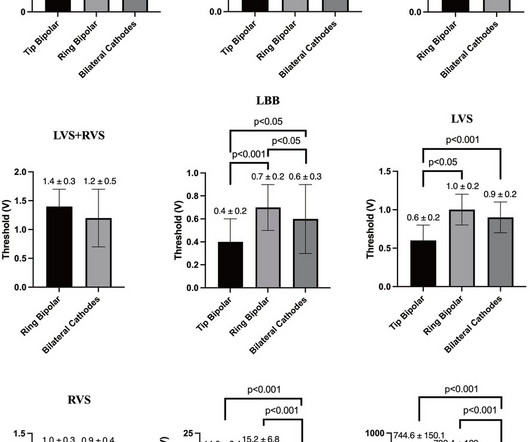
The electrocardiogram (ECG) and electrogram (EGM) parameters were evaluated and other electrophysiological characteristics were analyzed using a three-electrode configuration test.ResultsSeven capture modes [right ventricular septal (RVS)+left ventricular septal (LVS)+LBB, RVS+LBB, LVS+LBB, RVS+LVS, RVS, LVS, and LBB] were utilized in the study.
To diagnose heart conditions including heart attacks and heart rhythm disturbances, clinicians typically rely on 12-lead electrocardiograms (ECGs)—complex arrangements of electrodes and wires placed around the chest and limbs to detect the heart's electrical activity.
Our review study aimed to determine whether electrocardiogram (ECG) findings before PCI could serve as predictors for the occurrence of the no-reflow phenomenon. Methods and materials We systematically searched MEDLINE, Scopus, and Embase to identify relevant studies.
Initial diagnostic tools, such as the 12-lead electrocardiogram (ECG) and traditional ambulatory monitoring, frequently fall short in providing a conclusive diagnosis. Paroxysmal palpitations affect approximately 1 in 300 individuals, posing a significant challenge in correlating symptoms with cardiac rhythm disturbances.1
Notwithstanding many insightful observations, the electrocardiogram (ECG) arguably ignited the big bang in our understanding of cardiac arrhythmias. Using ECG recording and deductive reasoning, our teachers and predecessors classified the bradycardias and tachycardias and proposed many mechanisms, subsequently proven to be correct.
Patients had routine 12-lead electrocardiograms (ECGs) regardless of presenting complaints. Data regarding AF screening in conflict countries emergency departments (ED) is lacking.MethodsWe included consecutive patients >40 years old who reported to the ED of a Syrian tertiary centre between July 2024 and September 2024.
One area of interest is the use of LLMs as an aid in the interpretation of medical data and images of electrocardiograms (ECGs). As large language models (LLM) have become popular, the use of artificial intelligence (AI) for medical diagnostics has been on the forefront of medical circles and the news.
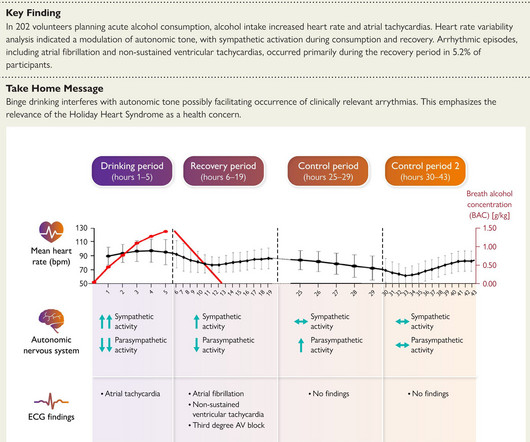
Medicine is subjecting the negative effects of alcohol on body and health to ever greater scrutiny. This should not surprise us, as alcohol is one of the strongest cell toxins that exist.
The electrocardiogram (ECG) had been used for the prediction of biological age (ECG-BA). Chronological age (CA) is a traditional powerful risk factor for aging-related diseases.
Artificial intelligence enhanced 12-lead electrocardiogram (AI-ECG) for detecting diastolic function and filling pressure has been developed. When patients present with dyspnea or shortness of breath in the emergency department (ED), it is important to promptly differentiate whether the symptoms are cardiac or extracardiac in origin.
Now, researchers and clinicians at Mayo Clinic are using artificial intelligence (AI) technology to flag heart problems earlier, boosting the abilities of a diagnostic test that has been around for over a century—the electrocardiogram (ECG).
Background: Characteristic electrocardiogram (ECG) changes in patients with Brugada-type ECG are augmented by a large meal, called “the full stomach test”, which has been reported to be associated with a history of life-threatening events.
Worldwide, over 300 million electrocardiograms (EKGs) are performed each year, with one-third of those taking place in the United States. Despite being so widely used, the technology of EKGs has been unchanged for decades.
Subsequent assessment revealed TTS. After receiving the optimal medical therapy, the patient was discharged after 10days without experiencing acute chest pain or shortness of breath.
The initial electrocardiogram showed type A preexcitation syndrome, with obvious ST-segment depression in leads V3 through V5 and positive delta wave. A man in his mid-50s presented with chest pain lasting 30 minutes. What would you do next?
Background Artificial intelligence (AI) has shown promise in the early detection of various cardiac conditions from a standard 12-lead electrocardiogram (ECG). However, the ability of AI to identify abnormalities from single-lead recordings across a range of pathological conditions remains to be systematically investigated.
VTT Technical Research Centre of Finland has developed a new sustainable electrocardiogram (ECG, also known as EKG) patch that is fully recyclable and made of biomaterials. The device is modular, so electronic components can be easily removed from the disposable patch and used again.
The ability to record an electrocardiogram (ECG) with a smartwatch (readily available without medical prescription), is a small revolution for cardiology.1 1 It allows individuals to play a more active role in their health care, improving understanding of cardiac function and its pathologies among individuals with and without cardiac disease.2
Convolutional neural networks (CNNs) have been used to build atrial fibrillation (AF) prediction models based on analysis of a sinus rhythm 12-lead electrocardiogram (ECG) using large datasets. Such datasets are not readily available for all clinical scenarios or diseases.
The first study, titled “Detection of Hypertrophic Cardiomyopathy on Electrocardiogram using Artificial Intelligence”, evaluated the performance of the Viz HCM algorithm across racial backgrounds at Mass General Hospital , Brigham and Women’s Hospital and Salem Hospital. “The sensitivity.
We organize all of the trending information in your field so you don't have to. Join thousands of users and stay up to date on the latest articles your peers are reading.
You know about us, now we want to get to know you!
Let's personalize your content
Let's get even more personalized
We recognize your account from another site in our network, please click 'Send Email' below to continue with verifying your account and setting a password.


Let's personalize your content