This site uses cookies to improve your experience. To help us insure we adhere to various privacy regulations, please select your country/region of residence. If you do not select a country, we will assume you are from the United States. Select your Cookie Settings or view our Privacy Policy and Terms of Use.
Cookie Settings
Cookies and similar technologies are used on this website for proper function of the website, for tracking performance analytics and for marketing purposes. We and some of our third-party providers may use cookie data for various purposes. Please review the cookie settings below and choose your preference.
Used for the proper function of the website
Used for monitoring website traffic and interactions
Cookie Settings
Cookies and similar technologies are used on this website for proper function of the website, for tracking performance analytics and for marketing purposes. We and some of our third-party providers may use cookie data for various purposes. Please review the cookie settings below and choose your preference.
Strictly Necessary: Used for the proper function of the website
Performance/Analytics: Used for monitoring website traffic and interactions
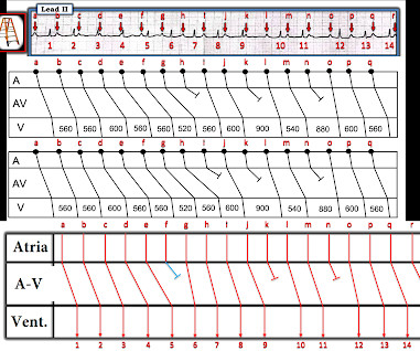
The long lead II rhythm strip shown in Figure-1 — was obtained from a previously healthy 30-year old woman, who presented with new abdominal pain. Her vital signs were stable — and she did not have an acute abdomen. QUESTIONS: Is there complete AV block? If not — How would YOU interpret this rhythm? What is unusual about this conduction disturbance?
Complex ECGs like this one have to be approached systematically. Firstly, we can see a normal sinus rhythm. A is the first beat of a wide complex tachycardia. This must be a ventricular tachycardia. Although there is a P-wave before the first beat of the tachycardia, it is not premature. Therefore, there is no SVT with aberrant conduction. The first beat of the tachycardia looks different from the subsequent beats because there is a fusion beat present.
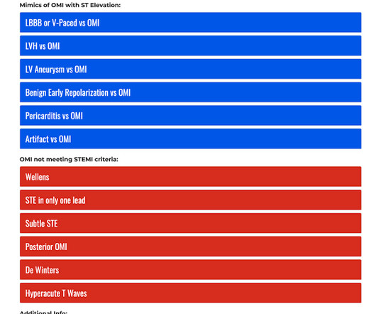
? OMI Pocket Guide The OMI Pocket Guide ( [link] ) is a user-friendly online resource designed to help healthcare professionals learn how to recognize subtle signs of acute coronary occlusion on the ECG which represent occlusion myocardial infarctions (OMI). Learning to recognize OMIs is an important clinical skill because it helps identify the subpopulation of "NSTEMIs" who are likely to be found with total thrombotic occlusion at the time of cardiac catherization.
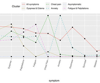
Objective This study aims to leverage natural language processing (NLP) and machine learning clustering analyses to (1) identify co-occurring symptoms of patients undergoing catheter ablation for atrial fibrillation (AF) and (2) describe clinical and sociodemographic correlates of symptom clusters. Methods We conducted a cross-sectional retrospective analysis using electronic health records data.
Speaker: Simran Kaur, Co-founder & CEO at Tattva Health Inc.
AI is transforming clinical trials—accelerating drug discovery, optimizing patient recruitment, and improving data analysis. But its impact goes far beyond research. As AI-driven innovation reshapes the clinical trial process, it’s also influencing broader healthcare trends, from personalized medicine to patient outcomes. Join this new webinar featuring Simran Kaur for an insightful discussion on what all of this means for the future of healthcare!
The ECG in Figure-1 was obtained from an 18-year old woman — who moments before been resuscitated from out-of-hospital cardiac arrest. QUESTIONS: In light of the above clinical history. How would YOU interpret her post-resuscitation ECG? Does this ECG in Figure-1 provide clue(s) to the etiology of this patient's cardiac arrest? Figure-1: The initial ECG in today's case — obtained following resuscitation from cardiac arrest of an 18-year old woman.
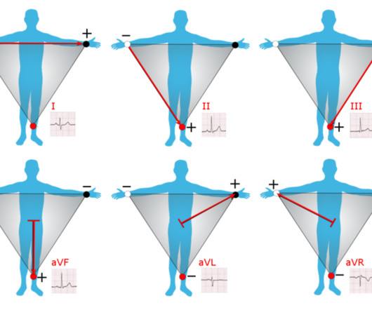
Introduction to ECG Testing & Einthoven’s Triangle: The electrocardiogram (ECG) represents the heart’s electrical activity, resulting from the contraction (depolarization) and relaxation (repolarization) of the atrial and ventricular muscles. When it comes to understanding the electrical activity of the heart, Einthoven’s Triangle is a fundamental concept in electrocardiography (ECG).
As a dedicated educator, you understand the challenges of providing your medical assistant students the best education. Limited resources and ever-evolving industry demands can often make it challenging to meet the high standards set by the healthcare field.
As a dedicated educator, you understand the challenges of providing your medical assistant students the best education. Limited resources and ever-evolving industry demands can often make it challenging to meet the high standards set by the healthcare field.
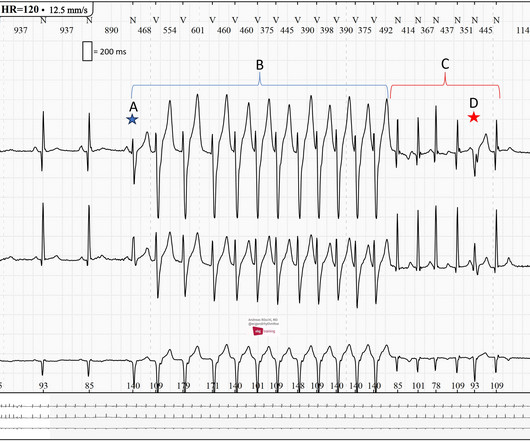
Here is a Pacemaker-ECG with no signs of PM-malfunction: Beat A is an intrinsic beat (atrial fibrillation). Beat B is a pseudofusion beat. Beat C is a fully paced beat. Beat D is a fusion beat. Ventricular fusion is the electrical summation of an intrinsic beat of the heart and depolarization from a pacing stimulus. The morphology lies between a fully paced beat and a complete intrinsic beat.
Rural hospitals across the United States are currently grappling with a severe and precarious financial situation. Over the past decade, more than 100 rural hospitals have shut their doors , leaving vulnerable communities without vital healthcare access. Alarmingly, these healthcare losses appear far from over, as an additional 600 rural hospitals, representing nearly 30% of all rural hospitals nationwide , face an imminent threat of closure.
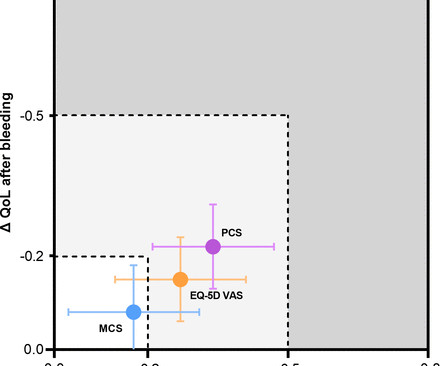
Objective Patients with acute coronary syndrome (ACS) remain at high risk for recurrent ischaemic and bleeding events during follow-up. Our study aimed to quantify and compare the impact of these adverse events on quality of life (QoL). Methods Data from patients with ACS prospectively enrolled in the FORCE-ACS registry between January 2015 and December 2019 were used for this study.
The healthcare industry continues to face a shortage of qualified providers. Hospital administrators have the unfathomably difficult job of maintaining sufficient healthcare staffing levels in a high-pressure environment with complex regulations and budget constraints.
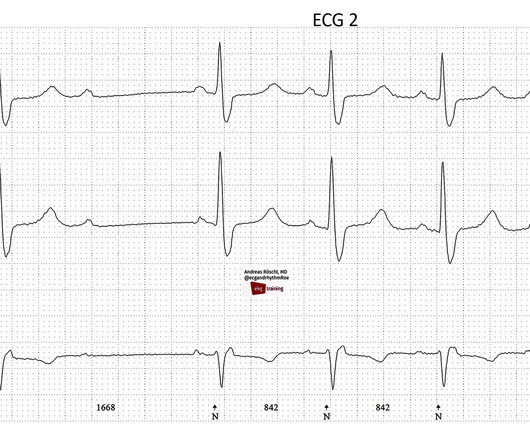
Why does this EKG indicate a sick sinus node? First, we observe a sinus rhythm with a rate just below 60 bpm. Then, there is a pause of approximately 3000 ms, followed not by a sinus beat, but by a junctional escape beat (retrograde/inverted P-wave immediately after the QRS complex). This ECG was recorded at the general practitioner's office, and it can be assumed that no vagal stimulus contributed to the arrhythmia.
Ever since the 12 lead ECG was introduced in the medical domain a key question is how to ensure anyone can interpret 12-lead ECG. With anyone we mean of course medical professionals but over the decades 12 lead ECG interpretation has remained a major challenge. Just imaging you have to read and interpret a set of 12 graphs each representing a different view on the heart during an activation and recovery cycle to understand what’s going on.
CT coronary angiography, in addition to a CT CAC, is arguably the best test for estimating whether someone has evidence of coronary artery disease and what that means for their near-term risk of a heart attack. This article is part 2 of a series on cardiac CT. Part one explored the utility and benefits of CT Calcium Scoring. If you have not yet read it, I suggest doing so before reading the remainder of this article.
At CTVS, our team of board-certified thoracic surgeons routinely perform a procedure known as a thoracic sympathectomy to treat hyperhidrosis, or excessive sweating. During this surgery, part of the sympathetic chain of nerves that runs along the spine is carefully severed to halt signals that trigger the sweat glands.
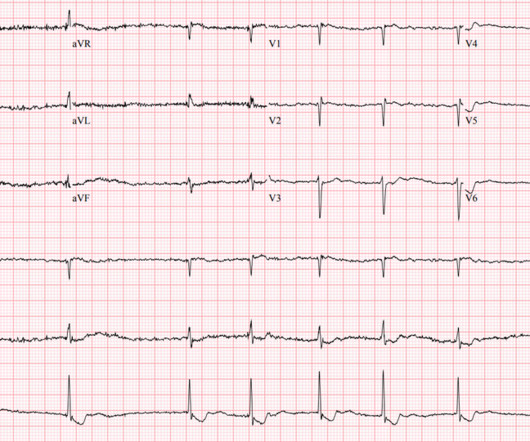
We are observing EKG strip 1 in a Holter EKG recording; what can be said about it? There is a sinus rhythm with a normal PQ interval. After 3 sinus beats, a 2:1 AV block develops. When 2:1 AV block occurs, we should not refer to this as Wenckebach (Mobitz I) or Mobitz II, but rather as a high-grade AV block (other forms include: 3:1, 4:1, 5:1, etc.).
Doctors, next time you’re seeing a freshly-diagnosed heart patient for a follow-up appointment, please remember that we’re far more than just a major organ that’s undergone a procedure.
David Didlake, FF/EMT-P, AG-ACNP @DidlakeDW An elder female presented to the ED with worsening shortness of breath. She was known to have a history of poorly controlled COPD, AFib, and multivessel coronary disease. Specific to the latter, she was previously deemed not appropriate for CABG (complex rationale) with preference for optimized medical management, instead.
Written by Pendell Meyers A woman in her 50s presented with acute chest pain and lightheadedness since the past several hours. Here is her triage ECG during active symptoms: What do you think? The ED physician read this as "Normal sinus rhythm. LVH. Marked ST abnormality, possible anterior subendocardial injury." Smith : I suspect this was a confirmation of the conventional computer interpretation.
A. Enrique Caballero, MD, will deliver the keynote address, Screening for Social Determinants of Cardiometabolic Health and Practice Implications, at the 18th Annual CMHC. Cardiometabolic Health Congress (CMHC) has announced details for the keynote address at its upcoming 18th annual conference, Social Determinants and Digital Advances in Cardiorenal Metabolic Health.
We are observing EKG strip 1 in a Holter EKG recording; what can be said about it? There is a sinus rhythm with a normal PQ interval. After 3 sinus beats, a 2:1 AV block develops. We should not refer to this as an AV block type Wenckebach (Mobitz I) or Mobitz II, but rather as a higher-degree AV block (other forms include: 3:1, 4:1, 5:1, etc.). The 2:1 block can be intranodally localized and behave benignly like a Wenckebach block typically does.
Immune checkpoint inhibitors (ICIs) are a form immunotherapy where the negative regulators of host immunity are targeted, thereby leveraging the own immune system. ICIs have significantly improved cancer survival in several advanced malignancies and there are currently over 90 different cancer indications for ICIs. The majority of patients develop immune-related adverse events (irAEs) during ICI therapy.
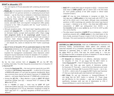
Broad complex tachycardia in a 78-year-old patient with coronary heart disease (CHD) and an old inferior myocardial infarction. Why is this a ventricular tachycardia (VT) and not a supraventricular tachycardia (SVT) with aberrant conduction? Broad complex tachycardia is generally about 80% likely to be ventricular in origin. However, in a patient with CHD and a history of myocardial infarction, this likelihood increases to about 90%.
The Patient: This ECG is from a 63-year-old man who complained of epigastric pain for three hours. The pain was sudden in onset, burning in nature, and accompanied by nausea and palpitations. The patient is a heavy smoker, diabetic and hypertensive with a long history of non-compliance to his medications. He was given crushed aspirin, loaded with clopidogrel and heparin, given high-intensity statins, and rushed to the cath lab.
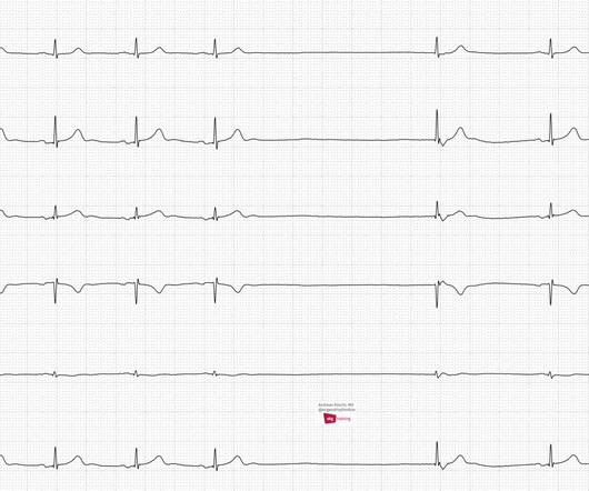
Here is the ECG of a 3-year-old boy. Is there cause for concern? The ECG shows a sinus rhythm with significant sinus arrhythmia. The heart rate increases with inspiration and decreases with expiration, which is called respiratory sinus arrhythmia. The QRS-axis is between 60 and 90 degrees, which is physiological at this age. The negative T-waves in V1-V3 (V4) are also age-appropriately normal.
We organize all of the trending information in your field so you don't have to. Join thousands of users and stay up to date on the latest articles your peers are reading.
You know about us, now we want to get to know you!
Let's personalize your content
Let's get even more personalized
We recognize your account from another site in our network, please click 'Send Email' below to continue with verifying your account and setting a password.


Let's personalize your content