This site uses cookies to improve your experience. To help us insure we adhere to various privacy regulations, please select your country/region of residence. If you do not select a country, we will assume you are from the United States. Select your Cookie Settings or view our Privacy Policy and Terms of Use.
Cookie Settings
Cookies and similar technologies are used on this website for proper function of the website, for tracking performance analytics and for marketing purposes. We and some of our third-party providers may use cookie data for various purposes. Please review the cookie settings below and choose your preference.
Used for the proper function of the website
Used for monitoring website traffic and interactions
Cookie Settings
Cookies and similar technologies are used on this website for proper function of the website, for tracking performance analytics and for marketing purposes. We and some of our third-party providers may use cookie data for various purposes. Please review the cookie settings below and choose your preference.
Strictly Necessary: Used for the proper function of the website
Performance/Analytics: Used for monitoring website traffic and interactions
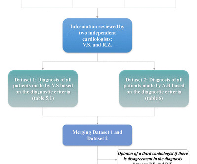
Background Determining heart failure (HF) phenotypes in routine electronic health records (EHR) is challenging. We aimed to develop and validate EHR algorithms for identification of specific HF phenotypes, using Read codes in combination with selected patient characteristics. Methods We used The Healthcare Improvement Network (THIN). The study population included a random sample of individuals with HF diagnostic codes (HF with reduced ejection fraction (HFrEF), HF with preserved ejection fractio
Case submitted and written by Mazen El-Baba MD, with edits from Jesse McLaren and edits/comments by Smith and Grauer A 90-year old with a past medical history of atrial fibrillation, type-2 diabetes, hypertension, dyslipidemia, presented with acute onset chest/epigastric pain, nausea, and vomiting. BP was 110 and oxygen saturation was normal. What is your ECG interpretation and what would you do next?
Change is one of the only constants in our lives, and we can count on it daily. While we might see change as a stressor, can you imagine your life without change? No improved health, no increase in pay, no new home, and no upcoming vacation!
30th October 2022 [How fewer doctors means more doctors – it’s official] This blog has nothing to do with heart disease, or vaccines, or anything directly about medical practice at all. However, it does have a great deal to do with data manipulation, which is something very close to my heart. It also illustrates how a ‘fact’ can be anything but. I am also hoping to help highlight an increasingly worrying trend that now scours the planet.
Speaker: Simran Kaur, Co-founder & CEO at Tattva Health Inc.
AI is transforming clinical trials—accelerating drug discovery, optimizing patient recruitment, and improving data analysis. But its impact goes far beyond research. As AI-driven innovation reshapes the clinical trial process, it’s also influencing broader healthcare trends, from personalized medicine to patient outcomes. Join this new webinar featuring Simran Kaur for an insightful discussion on what all of this means for the future of healthcare!
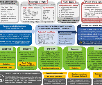
Introduction The diagnostic and therapeutic arsenal for heart failure with preserved ejection (HFpEF) has expanded. With novel therapies (eg, sodium-glucose co-transporter 2 inhibitors) and firmer recommendations to optimise non-cardiac comorbidities, it is unclear if outpatient HFpEF models can adequately deliver this. We; therefore, evaluated the efficacy of an existing dedicated HFpEF clinic to find innovative ways to design a more comprehensive model tailored to the modern era of HFpEF.
I was reading EKGs on the system and saw this one. What did I put in as my interpretation? Interpretation : "Acute LAD occlusion until proven otherwise. " There is non-diagnostic ST Elevation in V1-V3, with rather large T-waves but in the context of a deep S-wave (high voltage). HOWEVER, lead V4 is diagnostic of OMI. This is massive ST Elevation, huge hyperacute T-wave, and loss of S-wave (which in V4, unlike V2-3, can be normal but should greatly raise suspicion.
We organize all of the trending information in your field so you don't have to. Join thousands of users and stay up to date on the latest articles your peers are reading.
You know about us, now we want to get to know you!
Let's personalize your content
Let's get even more personalized
We recognize your account from another site in our network, please click 'Send Email' below to continue with verifying your account and setting a password.


Let's personalize your content