Fri.Sep 22, 2023

article thumbnail

ECG Blog #396 — Why the Flat Line?

Ken Grauer, MD

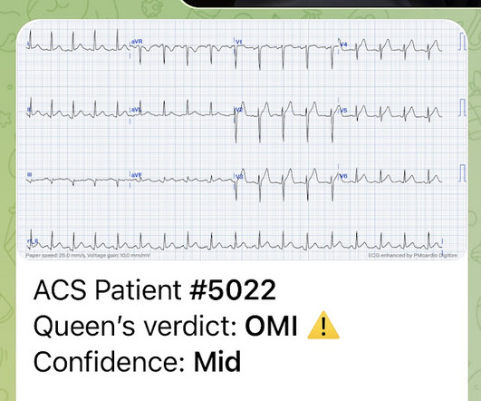
The ECG in Figure-1 — was obtained from a middle-aged man with palpitations and shortness of breath. He was hemodynamically stable at the time this tracing was recorded. How would YOU interpret the ECG in Figure-1 ? Is there evidence of a recent or ongoing acute MI? What might you do first? Figure-1: The initial ECG in today's case. ( To improve visualization — I've digitized the original ECG using PMcardio ).

Blog 168
article thumbnail

This ECG was texted to me: normal variant early repolarization, or LAD Occlusion MI (OMI)?

Dr. Smith's ECG Blog

This ECG was texted to me with no other information. I assumed the presentation was consistent with acute MI. What did I say? "OMI. Activate the cath lab." The T-waves in V2-V6 are diagnostic. It does, in fact, the STE meets STEMI criteria since there is 1 mm of in V4 and V5. There is also some non-diagnostic STE in inferior leads. There is zero reciprocal ST depression. 50% of LAD OMI have zero reciprocal ST depression!

STEMI 123
article thumbnail

Same genes behind heart muscle disorders in humans and Dobermanns

Science Daily - Heart Disease

Researchers have made a significant finding in determining the genetic background of dilated cardiomyopathy in Dobermanns. This research helps us understand the genetic risk factors related to fatal diseases of the heart muscle and the mechanisms underlying the disease, and offers new tools for their prevention.