Sat.Aug 26, 2023

article thumbnail

Instructors' Collection ECG - Inferior Posterior Wall M.I. In Cabrera Format

ECG Guru

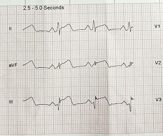
Does something about this ECG look "different" to you? This ECG shows a “classic” presentation of inferior-posterior M.I. when it is caused by a lesion in the right coronary artery (RCA). There are ST elevations in leads II, III, and aVF. Reciprocal ST depression is seen in Leads I and aVL. There is also reciprocal ST depression in Leads V1 – V3.

98
article thumbnail

Safety of Switching from a Vitamin K Antagonist to a Non-Vitamin K Antagonist Oral Anticoagulant in Frail Older Patients with Atrial Fibrillation: Results of the FRAIL-AF Randomized Controlled Trial

Circulation

Circulation, Ahead of Print. Background:There is ambiguity whether frail patients with atrial fibrillation (AF) managed with vitamin K antagonists (VKAs) should be switched to a non-vitamin K oral anticoagulant (NOAC).Methods:We conducted a pragmatic, multicenter, open-label, randomized controlled superiority trial. Older AF patients living with frailty (age ≥75 years plus a Groningen Frailty Indicator (GFI) score ≥3) were randomized to switch from INR-guided VKA treatment to a NOAC or to contin

article thumbnail

ECG Blog #392 — Repolarization T Waves?

Ken Grauer, MD

The ECG in Figure-1 was obtained from a man in his 60s — who described the sudden onset of "chest tightness" that began 20 minutes earlier, but who now ( at the time this ECG was recorded ) — was no longer having symptoms. In view of this history — How would YOU interpret this ECG? Should the cath lab be activated? Figure-1: The initial ECG in today's case. ( To improve visualization — I've digitized the original ECG using PMcardio ).

Blog 127