This site uses cookies to improve your experience. To help us insure we adhere to various privacy regulations, please select your country/region of residence. If you do not select a country, we will assume you are from the United States. Select your Cookie Settings or view our Privacy Policy and Terms of Use.
Cookie Settings
Cookies and similar technologies are used on this website for proper function of the website, for tracking performance analytics and for marketing purposes. We and some of our third-party providers may use cookie data for various purposes. Please review the cookie settings below and choose your preference.
Used for the proper function of the website
Used for monitoring website traffic and interactions
Cookie Settings
Cookies and similar technologies are used on this website for proper function of the website, for tracking performance analytics and for marketing purposes. We and some of our third-party providers may use cookie data for various purposes. Please review the cookie settings below and choose your preference.
Strictly Necessary: Used for the proper function of the website
Performance/Analytics: Used for monitoring website traffic and interactions
The work builds upon research published last year showing that an AI program can detect disease in the hearts mitral valve by analyzing ultrasound images of the heart. By applying AI to echocardiograms, we can help clinicians more easily detect the signs of heart valve disease so that patients get the care they need as soon as possible.
This clears the way for faster collaboration with other ultrasound systems, so the technology can reach more patients sooner. Weve proven that AI can empower novices to reliably perform echocardiograms, a breakthrough that could reshape how we diagnose and treat heart disease.
Food and Drug Adminstration (FDA) has approved DEFINITY (Perflutren Lipid Microsphere) as an ultrasound enhancing agent for use in pediatric patients with suboptimal echocardiograms, including those who have undergone heart transplant, or have Kawasaki disease or a congenital cardiovascular anomaly. Lantheus announced that the U.S.
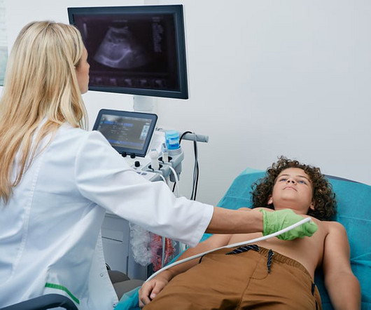
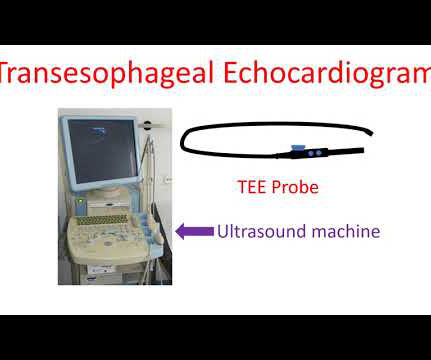
Echocardiogram is an image of the heart using ultrasound. An ultrasound beam is transmitted into the body using a device known as transducer. Transesophageal echocardiogram or TEE test, is obtained by introducing a special type of transducer, also called a TEE probe, through the throat into the food pipe (esophagus) and stomach.
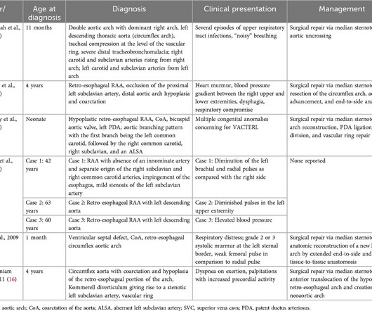
A 33-year-old G4P3 patient underwent fetal echocardiography after obstetric ultrasound showed concern for double aortic arch. Fetal echocardiogram was suspicious for vascular ring with presumptive diagnosis of double aortic arch vs. circumflex right aortic arch.
Artificial intelligence experts at Cedars-Sinai and the Smidt Heart Institute created a dataset with more than 1 million echocardiograms, or cardiac ultrasound videos, and their corresponding clinical interpretations.
Cedars-Sinai and Smidt Heart Institute investigators developed a novel foundation model that integrates computer vision interpretation of echocardiogram images with natural language processing to augment cardiologists’ interpretation of echocardiograms. Image by Getty.
11, 2025 UltraSight, a company committed to enhancing the efficiency and productivity of cardiac ultrasound,recentlyannounced support from Bristol Myers Squibb (BMS) for a study that aims to improve access to echocardiographic assessments for patients with obstructive hypertrophic cardiomyopathy (oHCM). tim.hodson Tue, 02/18/2025 - 16:17 Feb.
"Discover the latest guidelines from the European Society of Cardiology for managing chronic coronary syndromes, including the strong recommendation for using u
This case was posted on the [link] ultrasound site, of which this ECG blog is a part. I refer you to the video case presentation by one of my colleagues, Dr. Rob Reardon (who has, by the way, a fantastic collection of ED ultrasound cases). However, only the first ECG was shown, and it was recorded before the patient became ill.
This is the world’s only AI platform built on the world’s largest database of echocardiograms linked to mortality. One of our partners, Echo IQ, recently sought FDA approval for their AI-based cardiology workflow, EchoSolv.
A study featuring Fujifilm’s OPIE Transducer highlights that the system provides established imaging to aid in septal myectomy procedures compared to TEE and transthoracic echocardiogram (TTE). The OPIE transducer is compatible with Fujifilm’s premium ultrasound system, the ARIETTA Precision.
What do you think the echocardiogram shows? This was a point of care ultrasound, not a bubble contrast echo. Cath lab activated Dual antiplatelet therapy and heparin given. NTG drip started. Pain better still. First trop I returns at 1.5. POCUS Echo: POCUS Echo with no wall motion abnormality and normal ejection fraction.
The algorithm uses deep learning to analyse routine ultrasound scans of the heart ( echocardiograms ) to detect disease that often goes undetected during standard assessments.
Food and Drug Administration (FDA) has granted 510(k) clearance for its first-of-a-kind, AI-powered AISAP CARDIO point-of-care ultrasound (POCUS) software platform. We know that structural heart disease and heart failure are the leading causes of hospitalization and morbidity in the U.S.
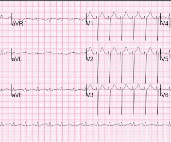
On arrival, lung ultrasound confirmed pulmonary edema (B lines). There is STE and hyperacute T-waves in V2 and V3, with significant STE in I and aVL, and inferior reciprocal STD. This is proximal LAD Occlusion until proven otherwise. An ECG was recorded: ED ECG 1: The findings are still present but not nearly as profound now.
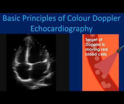
Usual colour Doppler echocardiogram is superimposition of colour Doppler images on a two dimensional echocardiogram. Colour M-Mode is superimposition of colour Doppler images on an M-Mode echocardiogram. Colour Doppler echocardiography receives the ultrasound signals reflected from moving red blood cells in the heart.
Bedside cardiac ultrasound showed moderately decreased LV function. EKG with paced complexes shown below shows much narrower QRS complex and echocardiogram showed improved LV systolic function primarily due to improvement in LV dyssynchrony. (J She was intubated. CT of the chest showed no pulmonary embolism but bibasilar infiltrates.
This comprehensive evaluation included the use of ultrasoundechocardiograms, computed tomography (CT) scans, electrocardiograms, mutagenesis analysis, and structural analysis to gain insights into the patient's condition and the underlying mechanisms of PD.
When following patients with serial echocardiograms, each new measur. Serial echocardiographic assessments are common in clinical cardiology, e.g., for timing of intervention in mitral and aortic regurgitation.
Ultrasound image of the heart – echocardiogram, showing fluid collection around the heart, marked as PE, short for pericardial effusion. Collection of fluid within the covering of the heart is called pericardial effusion. If it is severe enough to compress the heart, it prevents proper filling of the heart and blood pressure falls.
1.196 x STE60 in V3 in mm) + (0.059 x computerized QTc) - (0.326 x RA in V4 in mm) Third, one can do an immediate cardiac ultrasound. A bedside ultrasound was done by an emergency physician and simultaneously read by a cardiologist. greater than 23.4 is likely anterior STEMI). LV aneurysm is very different for inferior vs. anterior MI.
In this EM Quick Hits podcast we have Emily Austin on physostigmine for anticholinergic toxidrome, Walter Himmel on understanding nystagmus to differentiate central vs peripheral causes of vertigo, Rob Devins on the role of transesophageal echocardiogram in cardiac arrest, Jesse MacLaren on nuances in inferior MI ECG changes and aVL, Andrew Petrosoniak (..)
One very useful adjunct is ultrasound: Echo of his heart can distinguish aneurysm from acute MI by presence of diastolic dyskinesis, but it cannot distinguish demand ischemia from ACS. One must clearly rule out these processes before jumping on the ACS diagnosis. Furthermore, notice the well-formed Q-waves in inferior leads.
Hemodynamic instability in trauma is usually due to bleeding, but if ultrasound shows poor contractility, then this may be due to cardiac contusion. No further ECG, troponin, or echocardiogram was done because she was asymptomatic, and had a normal rhythm and rate. In the ED, ultrasound showed hemopericardium with tamponade.
Although he had a normal echocardiogram and stress test a year ago at a different hospital, due to his symptoms and intermediate-high risk probability of coronary artery disease (CAD), the decision was made to proceed with a cardiac catheterization using a trans-radial approach with a horizontal sweep technique.
The patient was thought to have low likelihood of ACS, and cardiology recommended repeat troponin, urine drug testing, and echocardiogram. Bedside echocardiogram showed hypokinesis of the mid to distal anterior wall and apex. Fortunately, this operator used intravascular ultrasound (IVUS). Initial hscTnI was 10 ng/L (ref. <14).
For example, by integrating Ventripoint’s AI-powered heart-scanning technology, which turns ultrasound images of the heart into MRI-quality heart images, InView provides pediatric cardiologists with access to MRI-quality heart images at a fraction of the cost and time needed for traditional MRIs. As well, by incorporating Us2.ai’s
Sometimes mild pericardial effusion may be detected by an echocardiogram done for other causes. Pericardial effusion is usually confirmed by an echocardiogram (ultrasound study of the heart). When the quantity is large enough to compress the heart, the person may feel breathless or dizzy because of a fall in blood pressure.
The patient underwent an emergent formal echocardiogram to look for wall motion abnormality: The estimated left ventricular ejection fraction is 63 %. Exclusion criteria were age less than 18, SBP less than 100 mmHg, echocardiogram with EF less than 50%, STEMI, pregnancy, and trauma. No wall motion abnormality.
During echocardiography, a transducer transmits the ultrasound beam towards the heart. The image shown here is an animated 2 dimensional echocardiogram. This one is an older mode known as time-motion mode or M-Mode echocardiogram. Hence a basic knowledge is needed for all physicians and paramedics.
Given her risk factors (HTN, HLD, ESRD from diabetes) I decided to obtain a broad cardiac workup for the patient: serial ECGs, labs, serial troponins, CXR and bedside cardiac ultrasound. This appears to be new, as her last formal echocardiogram 2 years ago was relatively normal. Clinical presentation is important, but so is history.
Two thirds of MINOCA cases are due to atherosclerotic causes One way to prove the diagnosis in this case would have been with intravascular imaging such as optical coherence tomography (OCT) or intravascular ultrasound (IVUS). His echocardiogram showed normal wall motion. Fortunately, that is exactly what happened.
He had diffuse crackles on exam and B-lines on chest ultrasound, and chest x-ray also confirmed pulmonary edema. Not all such ECGs represent anatomic aneurysms (on echo this is "diastolic dyskinesis"), but do generally represent an area of dense akinesis on echocardiogram. Blood pressure was 215/124 and HR 115 (on metoprolol).
So today i wanted to talk to you about what each heart test tells us about these different aspects of heart disease Tests that tell you about the heart as a pump The most commonly used test to assess the heart as a pump is an echocardiogram. This is an ultrasound (a bit like the type that we use on pregnant women to look at the baby).
This case was provided by Spencer Schwartz, an outstanding paramedic at Hennepin EMS who is on Hennepin EMS's specialized "P3" team, a team that receives extra training in advanced procedures such as RSI, thoracostomy, vasopressors, and prehospital ultrasound. This entire case is not consistent with takotsubo. It can only be seen by IVUS.
Troponins, echocardiogram An echocardiogram showed inferobasilar hypokinesis, further supporting a diagnosis of regional ischemia , likely of the area supplied by the RCA. Often, intravascular ultrasound or intravascular optical coherence tomography is requeried to make the diagnosis. The initial troponin I was elevated at 0.75
Smith comment: This patient did not have a bedside ultrasound. Had one been done, it would have shown a feature that is apparent on this ultrasound (however, this patient's LV function would not be as good as in this clip): This is recorded with the LV on the right. In fact, bedside ultrasound might even find severe aortic stenosis.
Here are a couple shots with strain, or "speckle tracking" on ED Echo: To, me these look like anterior wall motion abnormality, but I showed them to one of our ultrasound fellows who is very interested in this. They read it as normal. She said: This is a tough one.
Cupid EHR from Epic boasts the following: Cloud-based EHR Offers integrated order entry, scheduling, procedure documentation, structured reporting, and data analytics for cardiology practices Supports a wide range of workflows, including Echocardiograms, Ultrasound vascular, Cardiac Cath, stress testing, Electrophysiology, and structured documentation (..)
See this case: what do you think the echocardiogram shows in this case? Widespread ST-depression with reciprocal aVR ST-elevation can be cause by: Heart rate related: tachyarrhythmia (e.g., A emergent cardiology consult can be helpful for equivocal cases. POCUS showed good LV-function and no pericardial effusion.
The next morning the patient went for his routine echocardiogram, where the operator noticed a dilated aortic root at 5.47 Beware a negative Bedside ultrasound. Here is a quote from his initial cardiology admission note (after cath was done showing no acute culprit): ".chest Troponins gradually trended down from 0.19 Pericarditis?
ALL TROPS WERE UNDETECTABLE A formal ultrasound was done: Normal estimated left ventricular ejection fraction at rest. Next day, a stress echo was done: The exercise stress echocardiogram is normal. Normal estimated left ventricular ejection fraction improved with stress. No wall motion abnormality at rest.
Developed at Children’s National Hospital and detailed in the latest edition of the Journal of the American Heart Association , the new AI system combines the power of novel ultrasound probes with portable electronic devices installed with algorithms capable of diagnosing RHD on echocardiogram.
We organize all of the trending information in your field so you don't have to. Join thousands of users and stay up to date on the latest articles your peers are reading.
You know about us, now we want to get to know you!
Let's personalize your content
Let's get even more personalized
We recognize your account from another site in our network, please click 'Send Email' below to continue with verifying your account and setting a password.


Let's personalize your content