This site uses cookies to improve your experience. To help us insure we adhere to various privacy regulations, please select your country/region of residence. If you do not select a country, we will assume you are from the United States. Select your Cookie Settings or view our Privacy Policy and Terms of Use.
Cookie Settings
Cookies and similar technologies are used on this website for proper function of the website, for tracking performance analytics and for marketing purposes. We and some of our third-party providers may use cookie data for various purposes. Please review the cookie settings below and choose your preference.
Used for the proper function of the website
Used for monitoring website traffic and interactions
Cookie Settings
Cookies and similar technologies are used on this website for proper function of the website, for tracking performance analytics and for marketing purposes. We and some of our third-party providers may use cookie data for various purposes. Please review the cookie settings below and choose your preference.
Strictly Necessary: Used for the proper function of the website
Performance/Analytics: Used for monitoring website traffic and interactions
BackgroundElevated cardiac troponin (cTn) is detected in 10% to 30% of patients with acute ischemic stroke (AIS) and correlates with poor functional outcomes. Twenty‐two percent of patients with a rising pattern had an isolated dynamic cTn in the absence of any ECG or echocardiogram changes, compared with 53% with falling cTn.
The importance of transesophageal echocardiogram (TEE) to exclude left atrial (LA) and left atrial appendage (LAA) thrombus prior to cardioversion for atrial fibrillation (AF) has been debated in patients who are anticoagulated prior to cardioversion. Primary outcomes measured were the incidence of stroke within one-year post-cardioversion.
Today’s video is on the subject of stroke and in particular cryptogenic strokes. It is therefore crucial to identify the cause of stroke and treat the underlying cause wherever possible. Strokes are characterised by death of brain cells as a result of disruption of blood supply to those cells.
Stroke, Volume 55, Issue Suppl_1 , Page ATMP39-ATMP39, February 1, 2024. Patient management would be aided by inclusion of formal PFO shunt size quantification in all clinical stroke patient TEE reports. Results:Among the 50 patients, median age was 64 (IQR 58-74.5), and 20 (40%) were female.
A 90-something year old woman presented with an acute mild stroke. The diagnosis was a bit hard to find in the chart, and the echocardiogram did only stated "assymetric hypertrophy." She had a routine ECG as part of her workup: What do you think? This was shown to me in real time. But the patient had no symptoms.
The study’s findings, published in npj Digital Medicine , suggest AI could one day be employed to analyze images from a common imaging test called an echocardiogram, which uses sound waves to capture pictures of the heart. Left untreated, atrial fibrillation can cause stroke and heart failure. An estimated 12.1
Aim:This study investigates the prevalence of isolated interventricular membranous septal (IVMS) aneurysms detected via echocardiography and assesses the associated stroke risk without other classical risk factors.Methods:We searched the echocardiography database at Mount Sinai Morningside from January 2017 to September 2023.
Echocardiogram An echocardiogram uses sound waves to produce a detailed image of the heart, allowing doctors to see the size of the heart chambers and how well the heart is pumping blood. Blood Clots: An enlarged heart is more prone to developing blood clots, which can lead to stroke or pulmonary embolism.
While intracardiac cardiac tumors and shunts are infrequent and typically asymptomatic, their existence can precipitate severe outcomes, including stroke, myocardial infarction and sudden death.Case Description:A 69-year-old female presented with left sided facial droop, slurred speech and left arm weakness.
Stroke, Volume 56, Issue Suppl_1 , Page ATP182-ATP182, February 1, 2025. Advanced cardiac workup (ACW), including transesophageal echocardiogram (TEE) and implantable loop recorder (ILR) are widely considered a crucial element in the ESUS work-up. The etiology of AChA infarcts remains poorly understood.
Stroke, Volume 55, Issue Suppl_1 , Page ATP259-ATP259, February 1, 2024. Background and Purpose:Right-to-left shunt (RLS) is one of the potential embolic sources in embolic stroke of undetermined source (ESUS), but the eligibility of conducting shunt study to detect RLS in ESUS is still unknown. cm/s, p = 0.021).
Arrhythmias: Genetic mutations can also predispose individuals to irregular heart rhythms, such as atrial fibrillation or long QT syndrome, which may increase the risk of stroke or sudden cardiac arrest. Heart imaging, such as echocardiograms or CT scans. Key screenings include: Blood pressure and cholesterol checks.
Atrial fibrillation (AF) leads to impaired left atrial appendage contractility, increasing the risk of thromboembolic stroke. The left atrial appendage emptying velocity (LAAev) measured on transesophageal echocardiogram (TEE) is a marker of increased thromboembolic risk.
Transcript of the video: Closure line of aortic valve on M-Mode echocardiogram, is seen as central line, while in bicuspid aortic valve, it is an eccentric closure, nearer to one of the walls of the aorta. That is an important feature of bicuspid aortic valve on M-Mode echocardiogram. It forms almost like a box or rhomboid shape.
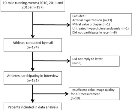
Methods Healthy, normotensive, male Caucasian participants of a 10-mile race were assessed with a 2D echocardiogram and comprehensive interview. Predictors of AD were investigated among training parameters by linear regression models corrected for age, resting heart rate, stroke volume index and mean blood pressure.
Stroke, Volume 56, Issue Suppl_1 , Page ATMP19-ATMP19, February 1, 2025. Background:Patients who arrive at the emergency department (ED) with transient ischemic attacks (TIA) are at an increased risk of experiencing a stroke. Radiology and cardiology have availability for urgent MRIs and echocardiograms.
BackgroundComplex aortic plaque (CAP) is a potential embolic source in patients with cryptogenic stroke (CS). Journal of the American Heart Association, Volume 12, Issue 23 , December 5, 2023.
Examples of cardio embolic stroke etiology include: 1. Atrial Fibrillation 2. Cardiomyopathy with mural thrombus 3. Patent Foramen Ovale 4. Severe calcific Aortic (valve) Stenosis 5. Mechanical prosthetic valve Severe carotid artery stenosis is also implicated in embolic stroke. Here is the admission ECG.
Stroke, Volume 55, Issue Suppl_1 , Page ATP274-ATP274, February 1, 2024. There is an elevated risk of thrombus formation and ischemic stroke in Rheumatic Heart Disease (RHD). The yearly prevalence of new stroke is 3-7.5% The yearly prevalence of new stroke is 3-7.5% But it is semi-invasive and have risks.
Compared to their peers without the condition, people with atrial fibrillation are twice as likely to be admitted to hospital, five times more likely to have a stroke, three times more likely to develop heart failure, and twice as likely to die prematurely.4 Symptoms include palpitations, fatigue, shortness of breath, dizziness, and fainting.
If these clots migrate to the blood vessels of the brain, a stroke may result. Ultrasound image of the heart – echocardiogram, showing fluid collection around the heart, marked as PE, short for pericardial effusion. Hence the blood stagnates in some parts of the upper chambers (left atrium) and clots may form.
The presence of type 2 diabetes not only signifies a chronic metabolic disorder, but also serves as a catalyst for various cardiovascular and cerebrovascular ailments such as coronary heart disease and stroke.
Stroke, Volume 56, Issue Suppl_1 , Page AWP44-AWP44, February 1, 2025. Background:Patent foramen ovale (PFO) is an independent risk factor for neurovascular injury such as stroke. Residual shunt post PFO closure was assessed using transthoracic echocardiogram (TTE) with saline contrast.
Brain MRI showed bilateral cortical and subcortical MCA ischemic strokes. TEE confirmed the recurrence of a left atrial myxoma, requiring a repeat sternotomy and resection.Since then, the patient was frequently hospitalized with seizures and strokes. Repeat echocardiograms did not show recurrence of a myxoma.
vs. 4.5%, p =0.96) on transesophageal echocardiogram did not differ. Both major (1.4% vs. 2.1%, p =0.72) and minor (27.8% vs. 19.4%, p =0.17) in-hospital complications were similar between the combined and control group, respectively. At 45 days, presence of peri-device leak (18.3% vs. 30.4%, p =0.07) and device related thrombosis (4.5%
Stroke: Vascular and Interventional Neurology, Volume 3, Issue S2 , November 1, 2023. He was admitted to the stroke service for work‐up of his abnormal brain vasculature. Remaining work‐up including A1c, LDL, urine drug screen, EKG, transthoracic echocardiogram, and telemetry was unrevealing other than for an LDL of 152.
A 35-year-old gravida 1, para 0 with biventricular heart failure (LVEF 25%), nonischemic cardiomyopathy, history stroke, history of left ventricular thrombus, class III obesity, and chronic kidney disease who had been followed by Cardio-Obstetrics throughout her pregnancy presented at 34 weeks gestation for planned induction of labor.
Important risks for cardiac catheterization in a deeply cyanotic infant are the chance of precipitation of a cyanotic spell and thrombotic strokes due to hemoconcentration. Another important role is for detection of coronary anomalies, which can also be seen on echocardiogram sometimes.
CTA head and neck were obtained and showed no evidence of intracranial hemorrhage, large vessel occlusion stroke (what a helpful and apt name for an acute arterial occlusion paradigm, by the way.), Echocardiogram was obtained and showed mild LVH without regional wall motion abnormality. Blood glucose was not low at 162 mg/dL.
All patients had an ejection fraction of 50% or higher as assessed with an echocardiogram performed within one week of their heart attack. There were also no differences in safety endpoints such as stroke, abnormally low blood pressure or fainting. Over a median follow-up period of 3.5 Over a median follow-up period of 3.5
In addition, AI’s ability to integrate and analyze diverse data sources—such as echocardiograms, MRI scans, and patient health records—provides a comprehensive view of a patient’s cardiovascular health. This holistic approach enhances diagnostic accuracy and supports more informed decision-making.
During aerobic exercise which is isotonic, the heart rate and stroke volume increases. Due to limitations of echocardiogram in evaluating the right ventricle, magnetic resonance imaging study of the right ventricle along with that of the left ventricle has been reported. Effect of exercise on right ventricle.
Echocardiogram showing thickened interventricular septum and mitral regurgitation in HCM. SAM causes LVOTO, increased ejection time and a decreased stroke volume, as well as mitral regurgitation due to poor coaptation of the leaflets. SAM in HCM Systolic anterior movement of mitral valve occurs in 30 – 60%, but it is not specific.
No further echocardiograms were available after cath. Free wall rupture, VSD, Dresslers Syndrome, chronic CHF, anatomic LV aneurysm, LV thrombus, stroke, etc). The patient was discharged one day after intervention and appears to be doing well. Teaching points: 1.
Although ApHCM has been considered a more “benign” variant, it is associated with increased risk of atrial and ventricular arrhythmias, apical thrombi, stroke, and progressive heart failure. Results In 414 patients, echocardiogram measurements of pulmonary artery systolic pressure (PASP) were obtained at the time of diagnosis.
Cardiac enzymes, CTs, echocardiograms, carotid ultrasounds, and electroencephalography all affected diagnosis or management in Postural blood pressure , performed in only 38% of episodes, had the highest yield with respect to affecting diagnosis (18-26%) or management (25-30%) and determining etiology of the syncopal episode (15-21%).
Written by Willy Frick A man in his 60s with hypertension and prior stroke presented with three days of crushing chest pain. Echocardiogram showed inferior wall hypokinesis. He reported intermittent chest pain for the last few months, but never lasting this long. He described it as substernal with radiation into the right arm.
We organize all of the trending information in your field so you don't have to. Join thousands of users and stay up to date on the latest articles your peers are reading.
You know about us, now we want to get to know you!
Let's personalize your content
Let's get even more personalized
We recognize your account from another site in our network, please click 'Send Email' below to continue with verifying your account and setting a password.


Let's personalize your content