This site uses cookies to improve your experience. To help us insure we adhere to various privacy regulations, please select your country/region of residence. If you do not select a country, we will assume you are from the United States. Select your Cookie Settings or view our Privacy Policy and Terms of Use.
Cookie Settings
Cookies and similar technologies are used on this website for proper function of the website, for tracking performance analytics and for marketing purposes. We and some of our third-party providers may use cookie data for various purposes. Please review the cookie settings below and choose your preference.
Used for the proper function of the website
Used for monitoring website traffic and interactions
Cookie Settings
Cookies and similar technologies are used on this website for proper function of the website, for tracking performance analytics and for marketing purposes. We and some of our third-party providers may use cookie data for various purposes. Please review the cookie settings below and choose your preference.
Strictly Necessary: Used for the proper function of the website
Performance/Analytics: Used for monitoring website traffic and interactions
In the setting of circumflex right aortic arch with the ductus arteriosus connecting the left descending aorta and left pulmonary artery, a vascular ring is present and can cause compressive symptoms of the aerodigestive tract. Fetal echocardiogram provides a unique opportunity to assess the aortic arch as the trachea is filled with fluid.
BackgroundDespite the poor outcomes related to the presence of pulmonary hypertension, it often goes undiagnosed in part because of low suspicion and screening tools not being easily accessible such as echocardiography. Each 15second PCG, recorded using a digital stethoscope, was processed to generate 5second melspectrograms.
She had acute pulmonary edema on exam. On arrival, lung ultrasound confirmed pulmonary edema (B lines). A 49 year old woman with h/o COPD only presented with sudden dyspnea. There is STE and hyperacute T-waves in V2 and V3, with significant STE in I and aVL, and inferior reciprocal STD.
link] Briefly, this woman without significant cardiac history went into pulmonary edema with respiratory failure. I refer you to the video case presentation by one of my colleagues, Dr. Rob Reardon (who has, by the way, a fantastic collection of ED ultrasound cases). In this case, the ECG never mimicked a STEMI.
Intubated and given nitric oxide for pulmonary hypertension. Echocardiogram during that time showed stiff pulmonic valve. Unlike adult hearts, the right ventricle comparatively large due to the work it has to do to pump against the high pulmonary pressure before birth. Weaned in NICU over 10 days.
He was started on a heparin drip and CTA of the chest was ordered to rule out pulmonary embolism. Echocardiogram showed severe RV dilation with McConnell’s sign and an elevated RVSP. Electrocardiographic Differentiation Between Acute Pulmonary Embolism and Acute Coronary Syndromes on the Basis of Negative T Waves - ScienceDirect.
Chronic Pulmonary Disease Lung diseases like chronic obstructive pulmonary disease (COPD) can lead to pulmonary hypertension, which in turn can cause the right side of the heart to enlarge, a condition known as cor pulmonale. The following diagnostic tools are commonly used: 1.
Clinical introduction A patient in their 30s had been diagnosed with peripartum cardiomyopathy, pulmonary oedema, with severe left ventricular dysfunction at the seventh month of gestation in the third pregnancy in their late 20s. Echocardiogram, CT aortogram and late gadolinium imaging of the aorta have been shown in figure 1.
He was in acute distress from pulmonary edema, with a BP of 180/110, pulse 110. He had diffuse crackles on exam and B-lines on chest ultrasound, and chest x-ray also confirmed pulmonary edema. The hypertension alone is the likely etiology of the pulmonary edema. He had no chest pain. The cath lab was activated.
A transthoracic echocardiogram demonstrated a large left atrial mass extending into the right upper pulmonary veins. A 73-year-old woman presented to the emergency department with a syncopal episode and a history of dizzy spells.
Animal studies have shown that mice with TBX1 gene mutations have smaller left pulmonary arteries compared to wild type mice, defined by a reduced left pulmonary artery (LPA) to right pulmonary artery (RPA) ratio. 0.27, p=0.002) echocardiogram. 0.18; p<0.001) and most recent (0.76±0.17
On day 3 of hospitalization, he experienced a syncopal episode and had acute worsening of hypoxemia that prompted a CT angiography of the chest which revealed bilateral, large clot burden pulmonary emboli with proximal thrombus in both the right and left main pulmonary arteries.
This is the schematic diagram of the heart in which you can see right atrium, right ventricle, left atrium, left ventricle, aorta and pulmonary artery. Similarly, another right sided valve is the pulmonary valve. There could also be some mild leak in the pulmonary valve, both these, in normal persons.
CT of the chest showed no pulmonary embolism but bibasilar infiltrates. EKG with paced complexes shown below shows much narrower QRS complex and echocardiogram showed improved LV systolic function primarily due to improvement in LV dyssynchrony. (J She was intubated. Bedside cardiac ultrasound showed moderately decreased LV function.
Xray was consistent with pulmonary vascular congestion. See this post: What do you think the echocardiogram shows in this case? Previously placed stents in the LAD (multiple) and mid circumflex and patent Formal echocardiogram: Normal left ventricular size and wall thickness. Lung exam showed diffuse B lines bilaterally.
Among these, a fistula between the left anterior descending artery and the pulmonary artery is the rarest variant, comprising about 17% of all coronary artery fistula cases.Case:A 54-year-old male, with a known history of atrial fibrillation and hypertension, presented to our emergency department with non-rotatory dizziness.
Transcript of the video: Closure line of aortic valve on M-Mode echocardiogram, is seen as central line, while in bicuspid aortic valve, it is an eccentric closure, nearer to one of the walls of the aorta. That is an important feature of bicuspid aortic valve on M-Mode echocardiogram. It forms almost like a box or rhomboid shape.
Tricuspid valve prolapse (TVP) can lead to TR and is sometimes overlooked, especially in complex cases with factors like pulmonary hypertension (PH). This treatment outcome and repeated echocardiograms reminded us that TR was primarily caused by TVP rather than PH alone.
Chance of precipitating a cyanotic spell are more when pulmonary angiography is attempted through the already narrow right ventricular outflow tract. Another important role is for detection of coronary anomalies, which can also be seen on echocardiogram sometimes. The image shows a MAPCA originating from right internal mammary artery.
This comprehensive evaluation included the use of ultrasound echocardiograms, computed tomography (CT) scans, electrocardiograms, mutagenesis analysis, and structural analysis to gain insights into the patient's condition and the underlying mechanisms of PD. Further genetic testing identified a homozygous mutation c.2662G>T
Advanced cardiac imaging especially in atypical presentations, can aid in early diagnosis.Case:A 59 year-old man with history of biopsy-proven pulmonary sarcoidosis presented with non exertional chest pain for 2 months. EKG, cardiac enzymes, and Initial echocardiogram(TTE) was unremarkable. The enhancement raised suspicion for CS.
This year's Boot Camp covered training in cardiopulmonary bypass skills, vessel anastomosis, diagnostic and therapeutic endoscopies, open pulmonary lobectomy, TAVR, and wire skills. The best parts of the Boot Camp were learning the basics of CT surgery, the vast topics covered (transthoracic echocardiogram, lobectomy, etc.)
CT Abdomen and Pelvis showed an enhancing heterogenous lesion at the left superior renal pole, left adrenal gland lesion, and a pulmonary artery filling defect. This defect, along with concern for metastasis, led to a CT chest angiogram that reported small left upper and lower lobe pulmonary emboli.
A pre-procedural transesophageal echocardiogram showed a left ventricular (LV) ejection fraction of 55%, mild concentric LV hypertrophy, and mild left atrial enlargement. A cardiac gated CT detailed the pulmonary vein structure and incidentally revealed bilateral cystic lung changes, leading to a pulmonology referral.
Methods Retrospective chart review of 200 patients admitted for ADHF from 2018 to 2019 with transthoracic echocardiogram during index hospitalisation. IVC diameter correlated to pulmonary arterial (PA) pressure (R=0.347, p<0.001) and body surface area (BSA) (R=0.424 p<0.001). Results The median age was 64, 30.5%
Objective To identify the most common transthoracic echocardiogram (TTE) parameters in patients hospitalised with severe acute respiratory syndrome coronavirus 2 (SARS-CoV-2/COVID-19) and their association with myocardial injury and outcomes. Remarkably, 90.8% had elevated troponin levels. to 7.94, p<0.03).
Similarly, for echocardiogram, what we would do usually is, first we do a clinical history evaluation, then physical examination, and after that only we proceed with echocardiography in our routine work. You can see the two dimensional sector imaging from an echocardiogram and I have marked out the aorta.
Afterward, a transesophageal echocardiogram guided implantation of both a Micra AV 2 (Medtronic) leadless pacemaker in the interventricular septum within the right ventricle and an Aveir (Abbott) leadless pacemaker in the superior base of the right atrial appendage was performed with successful pacing.
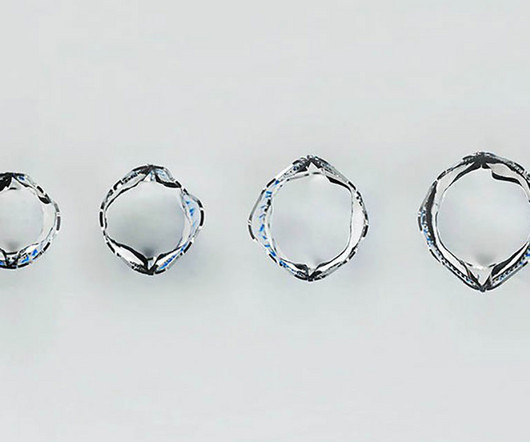
Unfortunately, the traditional pulmonary valves have a fixed diameter that can’t match the size of the child’s heart over time. The prototype valve has two leaflets crafted from a polymer material that has a well-established history of being used as a leaflet for pediatric pulmonary valves.
The Challenges Ascension Saint Thomas Hospital, based in Nashville, Tennessee, encountered persistent issues with missing documentation, including anesthesia records, Intraoperative Transesophageal Echocardiograms, perfusion records, and Pulmonary Function Test results.
After discharge, she was scheduled for a 2-week postpartum visit including echocardiogram, EKG, and NT-proBNP.Discussion:Given the patient's acute decompensation and fluid overload, medical optimization was essential prior to delivery. However, stabilization was expected to be temporary due to ongoing physiologic changes of pregnancy.
Echocardiogram is indicated (Correct) C. Start aspirin and Plavix Correct answer: (B) (B) Echocardiogram is indicated. Which of the following is the best statement to describe further clinical management? No further workup is indicated B. Start furosemide for diuresis D. Start with a Free Trial.
Hopefully a repeat echocardiogram will be performed outpatient. Q waves in association with RBBB are usually not seen in anterior leads unless there is pulmonary hypertension or anterior infarction. Systolic function normal by visual assessment only, unable to visualize well for further characterization. No cardiac MRI was done.
The image shown here is an animated 2 dimensional echocardiogram. This one is an older mode known as time-motion mode or M-Mode echocardiogram. The aorta, right ventricular outflow tract and pulmonary artery up to its bifurcation is imaged in the upward angulation shown in the left panel.
The patient underwent an emergent formal echocardiogram to look for wall motion abnormality: The estimated left ventricular ejection fraction is 63 %. Exclusion criteria were age less than 18, SBP less than 100 mmHg, echocardiogram with EF less than 50%, STEMI, pregnancy, and trauma. No wall motion abnormality.
First troponin I returns at 48 ng/L ECG 5 143 min No significant change ECG 6 261 min Same hs Troponin I profile (peaked at 1849): Formal Echocardiogram SUMMARY The estimated left ventricular ejection fraction is 74 %. The estimated pulmonary artery systolic pressure is 27 mmHg + RA pressure. Normal left ventricular cavity size.
The Challenges Ascension Saint Thomas Health, based in Nashville, Tennessee, encountered persistent issues with missing documentation, including anesthesia records, Intraoperative Transesophageal Echocardiograms (TEE), perfusion records, and Pulmonary Function Test (PFT) results.
He visited an outpatient clinic for it and an echocardiogram and exercise stress test was normal. On his physical examination, cardiac and pulmonary auscultation was completely normal. Bi-phasic scan showed no dissection or pulmonary embolism. He has 40 packs-year of smoking history. He denies taking any medication.
Pulmonary Arterial Hypertension (PAH): Hasan Ahmad Khasawneh’s (Jordan) systematic review of Treprostinil safety in PAH patients revealed no significant increase in mortality, though adverse events were more frequent. Memorial Lecture for Dr. Alain Cribier: Prof. Helene Eltchaninoff, MD (France) honored Dr.
Rupture can be either free wall rupture (causing tamonade) or septal rupture, causing ventricular septal defect with left to right flow and resulting pulmonary edema and shock. An echocardiogram showed no hemopericardium, but did show a new small ventricular septal defect with left to right shunting. 3) Oliva et al. (3)
Unfortunately there is no echocardiogram accessible because the patient checked himself out of the hospital in order to get back to his home state before it could be completed. A majority of patients with MAT have longstanding pulmonary disease. In the available view, the RCA appears fully occluded.
We organize all of the trending information in your field so you don't have to. Join thousands of users and stay up to date on the latest articles your peers are reading.
You know about us, now we want to get to know you!
Let's personalize your content
Let's get even more personalized
We recognize your account from another site in our network, please click 'Send Email' below to continue with verifying your account and setting a password.


Let's personalize your content