This site uses cookies to improve your experience. To help us insure we adhere to various privacy regulations, please select your country/region of residence. If you do not select a country, we will assume you are from the United States. Select your Cookie Settings or view our Privacy Policy and Terms of Use.
Cookie Settings
Cookies and similar technologies are used on this website for proper function of the website, for tracking performance analytics and for marketing purposes. We and some of our third-party providers may use cookie data for various purposes. Please review the cookie settings below and choose your preference.
Used for the proper function of the website
Used for monitoring website traffic and interactions
Cookie Settings
Cookies and similar technologies are used on this website for proper function of the website, for tracking performance analytics and for marketing purposes. We and some of our third-party providers may use cookie data for various purposes. Please review the cookie settings below and choose your preference.
Strictly Necessary: Used for the proper function of the website
Performance/Analytics: Used for monitoring website traffic and interactions
BackgroundComplex aortic plaque (CAP) is a potential embolic source in patients with cryptogenic stroke (CS). Most studies (74%) used ≥4 mm plaque thickness as the imaging criterion for CAP although ≥1 mm (N=1, CTA), ≥5 mm (N=5, TEE), and ≥6 mm (N=2, CTA) were also reported. 0.41],I2=94%). I2=95%) for TEE; 0.23 (95% CI, 0.15–0.34;I2=87%)
Mild Plaque no angiographically significant obstructive coronary artery disease. I looks as if there has been reperfusion. PM Cardio AI Bot: Not OMI with high confidence Angiogram: Speckle Tracking of Acute Pulm Edema.MOV from Stephen Smith on Vimeo.
The commonest causes of MINOCA include: atherosclerotic causes such as plaque rupture or erosion with spontaneous thrombolysis, and non-atherosclerotic causes such as coronary vasospasm (sometimes called variant angina or Prinzmetal's angina), coronary embolism or thrombosis, possibly microvascular dysfunction. It is not rare.
I interpreted the ECG as VT with two primary etiological possibilities: 1. Abrupt plaque ulceration of Type 1 ACS leading to VT. Corresponding echocardiogram demonstrated LV systolic dysfunction with an EF 30%. 2. Baseline fibrotic substrate from dilated cardiomyopathy leading to VT.
The patient was thought to have low likelihood of ACS, and cardiology recommended repeat troponin, urine drug testing, and echocardiogram. Bedside echocardiogram showed hypokinesis of the mid to distal anterior wall and apex. Smith comment : a very high proportion of MINOCA are ruptured plaque with lysed thrombus.
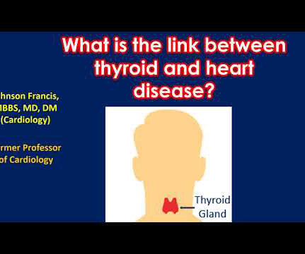
This in turn can enhance the chance of plaque build-up in the blood vessels of the heart (coronary arteries). Ultrasound image of the heart – echocardiogram, showing fluid collection around the heart, marked as PE, short for pericardial effusion. Reduced function of the thyroid gland is also associated with heart disease.
She underwent exercise echocardiogram in mid October where she exercised for nearly 7 minutes on the standard Bruce protocol and had typical anginal pain and shortness of breath. Baseline echocardiogram showed moderate LV systolic dysfunction with no wall motion abnormalities.
The scan did not find PE, but showed evidence of coronary plaque: There are areas of dense white in the LAD (red and blue circles) and in the first diagonal (green circle). An echocardiogram showed severely reduced global systolic function with an EF of 20-25% and an LV apical thrombus. An echocardiogram showed an EF of 20-25%.
Here is the cath report: Echocardiogram: There is severe hypokinesis of entire LV apex and apical segment of all the walls. To prove there is no plaque rupture, you need to do intravascular ultrasound (IVUS). An angiogram is a "lumenogram;" most plaque is EXTRALUMINAL!! ng/mL by 4th generation and older assays.)
Category 1 : Sudden narrowing of a coronary artery due to ACS (plaque rupture with thrombosis and/or downstream showering of platelet-fibrin aggregates. elevated BP), but rather directly correlated with coronary obstruction (due to plaque rupture and thrombosis) and, potentially, stymied TIMI flow. This results in Type I MI.
Troponins, echocardiogram An echocardiogram showed inferobasilar hypokinesis, further supporting a diagnosis of regional ischemia , likely of the area supplied by the RCA. Here’s the angiogram of the RCA : No thrombus or plaque rupture in the RCA (or any coronary artery) was found. The initial troponin I was elevated at 0.75
So today i wanted to talk to you about what each heart test tells us about these different aspects of heart disease Tests that tell you about the heart as a pump The most commonly used test to assess the heart as a pump is an echocardiogram. Overall though a normal cardiac MRI is even more reassuring than a normal echocardiogram.
Indeed, bedside Echocardiogram revealed severe left ventricular impairment of Takotsubo cardiomyopathy. The coronary angiogram revealed no critical stenosis, or acute plaque ulceration. Furthermore, pertinent electrolyte values (e.g. potassium) were within normal parameter.
She was treated medically for NonSTEMI, pending next day cath, which showed ulcerated plaque and a 60% thrombotic stenosis in the LAD distal to the first diagonal. A formal contrast echo was done at this point : Normal estimated left ventricular ejection fraction, 65%. Regional wall motion abnormality-distal septum and apex. It was stented.
See this case: what do you think the echocardiogram shows in this case? Widespread ST-depression with reciprocal aVR ST-elevation can be cause by: Heart rate related: tachyarrhythmia (e.g., POCUS showed good LV-function and no pericardial effusion. The patient had mild but diffuse abdominal tenderness.
Angiogram: Severe coronary artery calcification Moderate to severe distal small vessel disease mainly seen in RPL1, 2 Otherwise, Mild plaque, no angiographically significant obstructive coronary artery disease. Echocardiogram: Estimated left ventricular ejection fraction, lower limits of normal; 45-50%.
These are reperfusion T-waves (the same thing as Wellens' waves) Echocardiogram Regional wall motion abnormality-distal septum and apex. Regional wall motion abnormality-distal inferior wall. ECG recorded at time 38 hours: A further evolutionary stage of T-wave inversion.
Coronary Artery Disease (CAD) CAD, which involves the narrowing or blockage of coronary arteries due to plaque buildup, can reduce blood flow to the heart. Echocardiogram An echocardiogram uses sound waves to produce a detailed image of the heart, allowing doctors to see the size of the heart chambers and how well the heart is pumping blood.
Specific genetic variants, such as those affecting cholesterol metabolism, can increase the likelihood of plaque buildup in the arteries. Heart imaging, such as echocardiograms or CT scans. Cardiomyopathies: These diseases affect the heart muscle, impairing its ability to pump blood effectively.
Echocardiogram showed LVEF 66% with normal wall motion and normal diastolic function. His ECG at the accepting facility is shown below: Accepting facility ECG The team reviewed his angiography films with an interventionalist and thought they were suspicious for plaque rupture in LAD, but they were not confident.
With this test, an echocardiogram is done at rest to study the pumping ability of the heart. With time, fat and cholesterol can get trapped in the areas of wear and tear and cause plaque formation. The plaques can damage us in 2 ways.
However, an echocardiogram is a different test, also conducted for heart activity. Electrocardiogram, echocardiogram, and some other tests are done for patients with cardiac arrest. Coronary artery disease Excessive cholesterol builds up plaque that blocks the arteries supplying blood to the heart.
While awaiting transfer to the cath lab, STAT echocardiogram was performed and showed LVEF 30-35%, as well as anterior, inferior, and apical hypokinesis, and apical thrombus. As an aside, the LCx OMI is a type 2 event, since it is due to supply-demand mismatch from thrombus, and not due to atherosclerotic plaque rupture or erosion).
Nevertheless, the operator performed intravascular ultrasound and saw erupted calcium nodule consistent with plaque erosion. Echocardiogram showed inferior hypokinesis. As you can see, the lesion is not very angiographically impressive , more on this below. Troponin was rising when last checked, 8928 ng/L.
In this study of consecutive patients with LBBB who were hospitalized and had an echocardiogram, a QRS duration less than 170 ms (n = 262), vs. greater than 170 ms (n = 38), was associated with a significantly better ejection fraction (36% vs. 24%). So indeed the QRS is approximately 200 ms. Comment: What is the normal QRS duration in LBBB?
There were no plaques or stenoses. It is reasonable to perform an echocardiogram to evaluate LV function. A workup was undertaken in search of a cause of the patient's ventricular arrhythmia. As noted above echocardiography was completely normal. CT coronary angiogram showed a hypoplastic RCA and dominant LCx.
We organize all of the trending information in your field so you don't have to. Join thousands of users and stay up to date on the latest articles your peers are reading.
You know about us, now we want to get to know you!
Let's personalize your content
Let's get even more personalized
We recognize your account from another site in our network, please click 'Send Email' below to continue with verifying your account and setting a password.


Let's personalize your content