This site uses cookies to improve your experience. To help us insure we adhere to various privacy regulations, please select your country/region of residence. If you do not select a country, we will assume you are from the United States. Select your Cookie Settings or view our Privacy Policy and Terms of Use.
Cookie Settings
Cookies and similar technologies are used on this website for proper function of the website, for tracking performance analytics and for marketing purposes. We and some of our third-party providers may use cookie data for various purposes. Please review the cookie settings below and choose your preference.
Used for the proper function of the website
Used for monitoring website traffic and interactions
Cookie Settings
Cookies and similar technologies are used on this website for proper function of the website, for tracking performance analytics and for marketing purposes. We and some of our third-party providers may use cookie data for various purposes. Please review the cookie settings below and choose your preference.
Strictly Necessary: Used for the proper function of the website
Performance/Analytics: Used for monitoring website traffic and interactions
SCM may happen from a wide variety of psychological or physiological stresses, including respiratory failure (although in this case a psychological stress led to poor myocardial function and then pulmonary edema, then respiratory failure) and intracranial bleeding. The apparent trigger was stress from losing custody of children.
GradCAM++ technique highlighted physiologically meaningful PCG segments in example pulmonary hypertension recordings.ConclusionsWe demonstrated the feasibility of using digital stethoscopes in conjunction with deep learning algorithms as a lowcost, noninvasive, and easily accessible screening tool for early detection of pulmonary hypertension.
Introduction:The presence of a large physiologic shunt, defined as >20 left atrial microbubbles within 3 cardiac cycles on transesophageal echocardiography (TEE), is a key, randomized controlled trial-validated, indication for patent foramen ovale (PFO) with a closure device in patients with otherwise cryptogenic ischemic stroke.
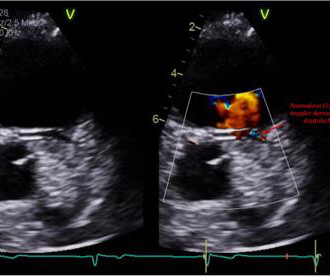
The echocardiogram showed normal cardiac structure and function, however, there was a concern for possible anomalous origin of the left coronary artery. A 14-year-old male with no significant medical history presented with intermittent palpitations for 2–3 months that occurred at rest and were associated with light-headedness.
Background:The emerging field of cardiac optogenetics uses pulses of light to stimulate optically-sensitive proteins in the heart, influencing cellular physiology with unprecedented spatiotemporal resolution. Circulation, Volume 150, Issue Suppl_1 , Page A4115027-A4115027, November 12, 2024.
After 5 weeks of the diet, HFD mice and their littermate controls underwent baseline exercise testing and echocardiograms. Echocardiograms were performed under 1.5% inhalational isoflurane. Gene and protein expression studies in heart tissue are underway. Further work is warranted with a larger sample size.
However, surgery was deferred because of decompensated HFpEF, as evidenced by significant leg edema on exam, and severely elevated left and right-side filling pressure, with restrictive physiology on right heart catheterization. LVOT obstruction resolved on follow up echocardiogram.
After discharge, she was scheduled for a 2-week postpartum visit including echocardiogram, EKG, and NT-proBNP.Discussion:Given the patient's acute decompensation and fluid overload, medical optimization was essential prior to delivery. However, stabilization was expected to be temporary due to ongoing physiologic changes of pregnancy.
It is a physiological adaptation helping athletes perform physical tasks better than non-athletes. Due to limitations of echocardiogram in evaluating the right ventricle, magnetic resonance imaging study of the right ventricle along with that of the left ventricle has been reported. Effect of exercise on right ventricle. References 1.Prior
ai’s echocardiogram algorithms, which automate measurements and pre-populate structured report templates, InView eliminates manual steps and improving the speed of coordination-of-care for patients with suspected heart disease. As well, by incorporating Us2.ai’s
But as the new variants and their implications started showcasing in the infected patients, it became clear that it has several physiological manifestations. Cardiovascular treatment reporting based on echocardiograms are needed to check the condition of the patients suffering from cardiovascular diseases.
So today i wanted to talk to you about what each heart test tells us about these different aspects of heart disease Tests that tell you about the heart as a pump The most commonly used test to assess the heart as a pump is an echocardiogram. Overall though a normal cardiac MRI is even more reassuring than a normal echocardiogram.
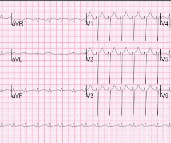
The combination of prolonged QT and deep T wave inversion throughout the precordium is typical of Takotsubo syndrome, or Stress Cardiomyopathy – which can occur in the context of a physiologically distressed ICU patient, further compromising their hemodynamics.
Get an emergent contrast echocardiogram. QTc's were 330 ms and 373 ms This is what I texted back: These look like they are a very pronounced case of Benign T-wave Inversion. I do not think this is acute occlusion myocardial infarction (OMI). These are reasons why it does not look like OMI: 1. flat ST segment in V4 2. huge R-wave in V4 3.
Next day echocardiogram showed inferolateral hypokinesia with an EF of %45-50. On echocardiogram you will not see a "posterior" hypokinesia (will see "inferolateral") and, as in this case, LCx may not give the blood supply of basal inferior segment (formerly called "posterior"). The patient recovered well.
This appears to be new, as her last formal echocardiogram 2 years ago was relatively normal. For some reason (with debatable physiology), coronary occlusion often causes decrease in the high voltage of LVH on the EKG. Parasternal Long Axis View There is a posterolateral wall motion abnormality.
A transthoracic echocardiogram showed an LV EF of less than 15%, critically severe aortic stenosis , severe LVH , and a small LV cavity. Any alteration in physiology can change "compensated" AS to "decompensated" AS. Inotropes and Vasopressors: Review of Physiology and Clinical Use in Cardiovascular Medicine. Circulation.
This T wave progression sequence does not make physiologic sense. By itself, this change in S wave appearance would not necessarily indicate lead malposition — BUT — the T wave becomes significantly taller from V1-to-V2 — then significantly smaller from V2-to-V3 — and then once again significantly taller from V3-to-V4.
Cardiac enzymes, CTs, echocardiograms, carotid ultrasounds, and electroencephalography all affected diagnosis or management in Postural blood pressure , performed in only 38% of episodes, had the highest yield with respect to affecting diagnosis (18-26%) or management (25-30%) and determining etiology of the syncopal episode (15-21%).
She needs an echocardiogram to rule out any subclinical myocardial disease. Echocardiogram is a must. Interpretation of T-wave inversion in physiological and pathological conditions: Current state and future perspectives. (Rarely, neuro-adrenergic-emotional signals from brainstem can tilt the ST segment like this. Reference 1.
We organize all of the trending information in your field so you don't have to. Join thousands of users and stay up to date on the latest articles your peers are reading.
You know about us, now we want to get to know you!
Let's personalize your content
Let's get even more personalized
We recognize your account from another site in our network, please click 'Send Email' below to continue with verifying your account and setting a password.


Let's personalize your content