This site uses cookies to improve your experience. To help us insure we adhere to various privacy regulations, please select your country/region of residence. If you do not select a country, we will assume you are from the United States. Select your Cookie Settings or view our Privacy Policy and Terms of Use.
Cookie Settings
Cookies and similar technologies are used on this website for proper function of the website, for tracking performance analytics and for marketing purposes. We and some of our third-party providers may use cookie data for various purposes. Please review the cookie settings below and choose your preference.
Used for the proper function of the website
Used for monitoring website traffic and interactions
Cookie Settings
Cookies and similar technologies are used on this website for proper function of the website, for tracking performance analytics and for marketing purposes. We and some of our third-party providers may use cookie data for various purposes. Please review the cookie settings below and choose your preference.
Strictly Necessary: Used for the proper function of the website
Performance/Analytics: Used for monitoring website traffic and interactions
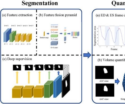
BackgroundPercutaneous extracorporeal membrane oxygenation (ECMO) is administered to pediatric patients with cardiogenic shock or cardiac arrest. Echocardiogram was performed for patients with ECMO, including at pre-ECMO, during cannulation, during ECMO support, during the ECMO wean, and a follow up within 3 months after weaning.
Echocardiogram during that time showed stiff pulmonic valve. The patient: 4 week old female infant with past medical history of meconium aspiration at birth with APGAR scores of 2,4,6. Intubated and given nitric oxide for pulmonary hypertension. Weaned in NICU over 10 days. This ECG was obtained at follow up appointment.
We report on the refinement and validation of a pediatric echocardiography complexity (PEC) score.Methods and ResultsThe American College of Cardiology Quality Network assembled a panel from 12 centers to refine a previously published PEC score developed in a single institution. Among the 174 echocardiograms analyzed, 68.9%
Introducing Sectra Echo Viewer Coming Spring 2025, the Sectra Echo Viewer will transform echocardiogram workflows with: Lightning-fast loading and crystal-clear playback Same full diagnostic quality in Sectra IDS7 and Sectra UniView Comprehensive measurement tools with structured reporting export Multi-protocol support for TTE, TEE, vascular, and pediatric (..)
Developed at Children’s National Hospital and detailed in the latest edition of the Journal of the American Heart Association , the new AI system combines the power of novel ultrasound probes with portable electronic devices installed with algorithms capable of diagnosing RHD on echocardiogram.
Changes in LVFS, LVEDD, LV end‐diastolic posterior wall thickness, and the LV end‐diastolic posterior wall thickness:LVEDD ratio between baseline and follow‐up echocardiograms acquired ≈1 year after diagnosis were determined for children who, at the 1‐year follow‐up had died, received a heart transplant, or were alive and transplant‐free.
Methods We performed a retrospective analysis to examine the clinical manifestations, genetic traits, and the relationship between PD and mitochondrial function in a pediatric patient.
For example, by integrating Ventripoint’s AI-powered heart-scanning technology, which turns ultrasound images of the heart into MRI-quality heart images, InView provides pediatric cardiologists with access to MRI-quality heart images at a fraction of the cost and time needed for traditional MRIs. As well, by incorporating Us2.ai’s
conducted extensive training using EchoNet-LVH, a primary dataset comprising 12,000 meticulously labeled echocardiogram videos sourced from diverse patients exhibiting various cardiac conditions. in the pediatric dataset and 5.6% This novel approach ensures high accuracy and enables real-time deployment in clinical settings.We
We believe they are likely a normal variant in this context, and the study above failed to identify any clinically significant finding after exam and echocardiogram in 110 children with bifid T waves. It is important to remember that pediatric tracings manifest a number of differences from adult ECGs.
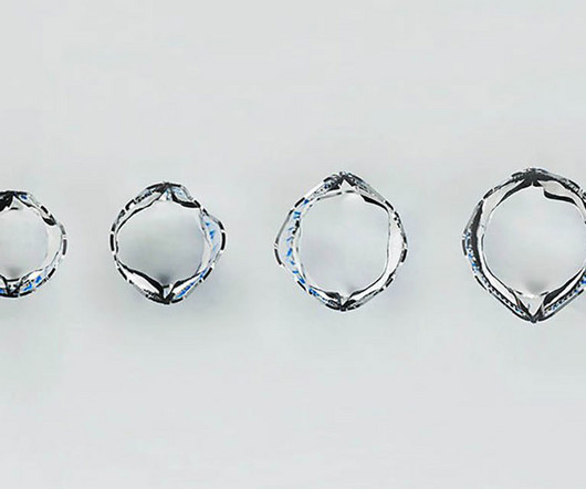
The prototype valve has two leaflets crafted from a polymer material that has a well-established history of being used as a leaflet for pediatric pulmonary valves. And following the valve implantation procedure, doctors assess the valve’s integrity and blood flow control through an echocardiogram.
The image shown here is an animated 2 dimensional echocardiogram. This one is an older mode known as time-motion mode or M-Mode echocardiogram. Subcostal view is a favourite view of pediatric echocardiographers. Unlike the previous 2 dimensional imaging, this is a single dimensional imaging.
No further ECG, troponin, or echocardiogram was done because she was asymptomatic, and had a normal rhythm and rate. After this case , were I to see a similar one, I would: 1) do a formal echocardiogram on anyone with new significant ECG abnormalities. She was discharged to home feeling just fine.
Formal echocardiogram showed normal EF, no wall motion abnormalities, no pericardial effusion. Pediatric and elderly patients were more predisposed to developing an arrhythmic event in the setting of fever [7]. Another troponin was drawn around the time of cath, troponin T (older generation), which was normal at less than 0.01
Food and Drug Adminstration (FDA) has approved DEFINITY (Perflutren Lipid Microsphere) as an ultrasound enhancing agent for use in pediatric patients with suboptimal echocardiograms, including those who have undergone heart transplant, or have Kawasaki disease or a congenital cardiovascular anomaly. Lantheus announced that the U.S.
To create comprehensive reports, connect to applications dealing with 4D, echocardiography, nuclear medicine, CT angiography, and pediatric echo reporting. This is a great way to increase productivity in any medical setting.
A formal echocardiogram was completed the next day and again showed a normal ejection fraction without any focal wall motion abnormalities to suggest CAD. Pediatric and elderly patients were more predisposed to developing an arrhythmic event in the setting of fever [7].
In this pediatric study, it was 71% successful and better than amiodarone. I have ordered an echocardiogram which will be done today, after that patient can be discharged to home with follow-up in 2 to 3 months." Procainamide is another reasonable solution to the problem. The echo was normal. Learning points 1.
Model performance was evaluated on single ECG–echocardiogram pairs per patient at Boston Children’s Hospital and externally at Mount Sinai Hospital using area under the receiver operating characteristic curve (AUROC) and area under the precision-recall curve (AUPRC).RESULTS:The composite outcomes) cohorts. and 99.2%, respectively.
We organize all of the trending information in your field so you don't have to. Join thousands of users and stay up to date on the latest articles your peers are reading.
You know about us, now we want to get to know you!
Let's personalize your content
Let's get even more personalized
We recognize your account from another site in our network, please click 'Send Email' below to continue with verifying your account and setting a password.


Let's personalize your content