This site uses cookies to improve your experience. To help us insure we adhere to various privacy regulations, please select your country/region of residence. If you do not select a country, we will assume you are from the United States. Select your Cookie Settings or view our Privacy Policy and Terms of Use.
Cookie Settings
Cookies and similar technologies are used on this website for proper function of the website, for tracking performance analytics and for marketing purposes. We and some of our third-party providers may use cookie data for various purposes. Please review the cookie settings below and choose your preference.
Used for the proper function of the website
Used for monitoring website traffic and interactions
Cookie Settings
Cookies and similar technologies are used on this website for proper function of the website, for tracking performance analytics and for marketing purposes. We and some of our third-party providers may use cookie data for various purposes. Please review the cookie settings below and choose your preference.
Strictly Necessary: Used for the proper function of the website
Performance/Analytics: Used for monitoring website traffic and interactions
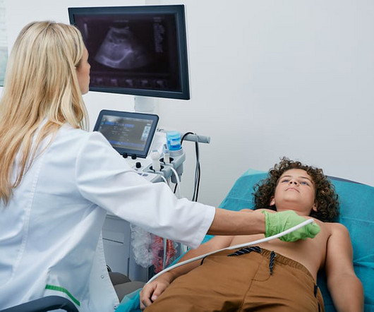
The work builds upon research published last year showing that an AI program can detect disease in the hearts mitral valve by analyzing ultrasound images of the heart. By applying AI to echocardiograms, we can help clinicians more easily detect the signs of heart valve disease so that patients get the care they need as soon as possible.
Food and Drug Adminstration (FDA) has approved DEFINITY (Perflutren Lipid Microsphere) as an ultrasound enhancing agent for use in pediatric patients with suboptimal echocardiograms, including those who have undergone heart transplant, or have Kawasaki disease or a congenital cardiovascular anomaly. Lantheus announced that the U.S.
Though the public at large currently pictures consumer-focused tools like ChatGPT and the Dall-E image generator when they think of AI, a myriad of AI-based applications are being implemented in the background of healthcare systems around the country, improving patient outcomes and enhancing the workflows of providers everywhere.
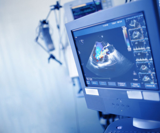
The algorithm uses deep learning to analyse routine ultrasound scans of the heart ( echocardiograms ) to detect disease that often goes undetected during standard assessments. Cardiac amyloidosis is a heterogeneous disease that results from the accumulation of abnormal proteins within the heart, impairing its ability to pump blood.
Food and Drug Administration (FDA) has granted 510(k) clearance for its first-of-a-kind, AI-powered AISAP CARDIO point-of-care ultrasound (POCUS) software platform. We know that structural heart disease and heart failure are the leading causes of hospitalization and morbidity in the U.S.
On arrival, lung ultrasound confirmed pulmonary edema (B lines). Outcome : She was diagnosed with stress cardiomyopathy, though it is not entirely classic. There is STE and hyperacute T-waves in V2 and V3, with significant STE in I and aVL, and inferior reciprocal STD. This is proximal LAD Occlusion until proven otherwise.
Hemodynamic instability in trauma is usually due to bleeding, but if ultrasound shows poor contractility, then this may be due to cardiac contusion. No further ECG, troponin, or echocardiogram was done because she was asymptomatic, and had a normal rhythm and rate. In the ED, ultrasound showed hemopericardium with tamponade.
Providing exceptional cardiovascular care for patients to achieve the best possible outcomes is the number one goal for cardiologists, hands-down. The best possible outcome of this situation is inefficient workflow, driving higher overall cost of care and resulting in widespread physician and patient frustration. Why is that?
Although he had a normal echocardiogram and stress test a year ago at a different hospital, due to his symptoms and intermediate-high risk probability of coronary artery disease (CAD), the decision was made to proceed with a cardiac catheterization using a trans-radial approach with a horizontal sweep technique.
He had diffuse crackles on exam and B-lines on chest ultrasound, and chest x-ray also confirmed pulmonary edema. Not all such ECGs represent anatomic aneurysms (on echo this is "diastolic dyskinesis"), but do generally represent an area of dense akinesis on echocardiogram. Blood pressure was 215/124 and HR 115 (on metoprolol).
Troponins, echocardiogram An echocardiogram showed inferobasilar hypokinesis, further supporting a diagnosis of regional ischemia , likely of the area supplied by the RCA. Often, intravascular ultrasound or intravascular optical coherence tomography is requeried to make the diagnosis. The initial troponin I was elevated at 0.75
This case was provided by Spencer Schwartz, an outstanding paramedic at Hennepin EMS who is on Hennepin EMS's specialized "P3" team, a team that receives extra training in advanced procedures such as RSI, thoracostomy, vasopressors, and prehospital ultrasound. This entire case is not consistent with takotsubo. It can only be seen by IVUS.
This enables early intervention, minimizes hospital admissions, and helps improve patient outcomes. OMS EHR features can improve chronic care management and principal care management, and they include: Offering cloud-based cardiology-focused EHR Accessing a color-coded dashboard that highlights those patients in need of attention first.
See this case: what do you think the echocardiogram shows in this case? As a result — cardiac cath was not performed — since results of a cath would not have altered the unfortunate outcome. Widespread ST-depression with reciprocal aVR ST-elevation can be cause by: Heart rate related: tachyarrhythmia (e.g.,
Developed at Children’s National Hospital and detailed in the latest edition of the Journal of the American Heart Association , the new AI system combines the power of novel ultrasound probes with portable electronic devices installed with algorithms capable of diagnosing RHD on echocardiogram.
and these are associated with larger MI and worse outcomes (2. If detected early by ultrasound, the patient can be saved. Our own Dave Plummer of HCMC reported on survival of 2 of 6 patients with free wall myocardial rupture diagnosed by bedside ultrasound in the ED.(3) Very unlikely. Raitt et al.) [and 3) Oliva et al. (3)
such Q-waves are associated with larger MI and worse outcomes (2. An echocardiogram showed no hemopericardium, but D oppler showed a new small ventricular septal defect with left to right shunting. If detected early by ultrasound, the patient can be saved. Very unlikely. Raitt et al.), Armstrong et al.), 3) Oliva et al. (4)
After rethinking the case, he remained concerned about ACS and subsequently performed a point-of-care ultrasound in order to evaluate for regional wall motion abnormality. He underwent formal echocardiogram several days later, which confirmed the findings of anterior, and apical wall motion abnormalities. Do NOT use them.
Case continued: All the physicians were very worried about LAD occlusion and recorded a couple bedside ultrasounds: This shows a profound apical and septal wall motion abnormality, perfectly consistent with LAD OMI. These ultrasounds confirm LAD occlusion. Journal of the American College of Cardiology 25 (5): 1084–88.
Check : [vitals, SOB, Chest Pain, Ultrasound] If the patient has Abdominal Pain, Chest Pain, Dyspnea or Hypoxemia, Headache, Hypotension , then these should be considered the primary chief complaint (not syncope). Cardiac Syncope ("True Syncope") Independent Predictors of Adverse Outcomes condensed from multiple studies 1.
Nevertheless, the operator performed intravascular ultrasound and saw erupted calcium nodule consistent with plaque erosion. Echocardiogram showed inferior hypokinesis. As you can see, the lesion is not very angiographically impressive , more on this below. Troponin was rising when last checked, 8928 ng/L.
I suspect pulmonary edema, but we are not given information on presence of B-lines on bedside ultrasound, or CXR findings. Severe AS markedly complicates management and portends poorer longterm outcome when associated with acute MI ( Abraham et al Cath Cardiovasc Interv 102(1): 159-165, 2023 Paquin et al Open Heart 9(1):e002046, 2022 ).
We organize all of the trending information in your field so you don't have to. Join thousands of users and stay up to date on the latest articles your peers are reading.
You know about us, now we want to get to know you!
Let's personalize your content
Let's get even more personalized
We recognize your account from another site in our network, please click 'Send Email' below to continue with verifying your account and setting a password.


Let's personalize your content