Remove Echocardiogram Remove Laboratory Remove Tachycardia
article thumbnail

Dyspnoea, dizziness and dysrhythmia in a middle-aged patient

Heart BMJ

ECG at presentation was suggestive of ventricular tachycardia (VT) ( figure 1 A ). Resuscitation with urgent cardioversion in view of haemodynamic instability with wide complex tachycardia was done. Laboratory evaluation revealed electrolytes, haematological profile, renal and liver panel to be within normal limits.

article thumbnail

Case Report: Comprehensive evaluation of ECG phenotypes and genotypes in a family with Brugada syndrome carrying SCN5A-R376H

Frontiers in Cardiovascular Medicine

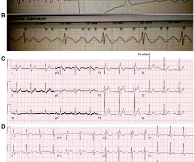
Patients with BrS can be asymptomatic or present with symptoms secondary to polymorphic ventricular tachycardia or ventricular fibrillation. The routine laboratory results, imaging study, coronary angiogram, and echocardiogram (ECG) were normal. The patient did not have underlying diseases.

article thumbnail

Chest Pain and Inferior ST Elevation.

Dr. Smith's ECG Blog

There is sinus tachycardia. Sinus tachycardia, which exaggerates ST segments and implies that there is another pathology. I have always said that tachycardia should argue against acute MI unless there is cardiogenic shock or 2 simultaneous pathologies. Here is that ECG: What do you think? No wall motion abnormality. Apple, Ph.D.

article thumbnail

Critical Left Main

EMS 12-Lead

Category 2 : An increase in myocardial oxygen demand due to tachycardia, elevated ventricular afterload (BP or aortic stenosis), or increased wall stretch (admittedly this latter is more complicated) or a decrease in oxygen supply due to hypotension, anemia, hypoxia, or a combination of all of the above. This results in Type I MI.

Angina 52
article thumbnail

Emergency Department Syncope Workup: After H and P, ECG is the Only Test Required for Every Patient.

Dr. Smith's ECG Blog

If the patient has Abnormal Vital Signs (fever, hypotension, tachycardia, or tachypnea, or hypoxemia), then these are the primary issue to address, as there is ongoing pathology which must be identified. Most physicians will automatically be worried about these symptoms. The tracings were considered abnormal in the following cases: 1.