This site uses cookies to improve your experience. To help us insure we adhere to various privacy regulations, please select your country/region of residence. If you do not select a country, we will assume you are from the United States. Select your Cookie Settings or view our Privacy Policy and Terms of Use.
Cookie Settings
Cookies and similar technologies are used on this website for proper function of the website, for tracking performance analytics and for marketing purposes. We and some of our third-party providers may use cookie data for various purposes. Please review the cookie settings below and choose your preference.
Used for the proper function of the website
Used for monitoring website traffic and interactions
Cookie Settings
Cookies and similar technologies are used on this website for proper function of the website, for tracking performance analytics and for marketing purposes. We and some of our third-party providers may use cookie data for various purposes. Please review the cookie settings below and choose your preference.
Strictly Necessary: Used for the proper function of the website
Performance/Analytics: Used for monitoring website traffic and interactions
Bedside cardiac ultrasound showed moderately decreased LV function. CASE CONTINUED She was admitted to the ICU. EKG with paced complexes shown below shows much narrower QRS complex and echocardiogram showed improved LV systolic function primarily due to improvement in LV dyssynchrony. (J She was intubated. J Am Coll Cardiol.
Course : A CT of the head, neck, chest, abdomen and pelvis showed no other unanticipated injuries and she was admitted to the ICU. Hemodynamic instability in trauma is usually due to bleeding, but if ultrasound shows poor contractility, then this may be due to cardiac contusion. She was discharged to home feeling just fine.
Given her risk factors (HTN, HLD, ESRD from diabetes) I decided to obtain a broad cardiac workup for the patient: serial ECGs, labs, serial troponins, CXR and bedside cardiac ultrasound. This appears to be new, as her last formal echocardiogram 2 years ago was relatively normal. Clinical presentation is important, but so is history.
The next morning the patient went for his routine echocardiogram, where the operator noticed a dilated aortic root at 5.47 in the ICU but survived with excellent function. Beware a negative Bedside ultrasound. Here is a quote from his initial cardiology admission note (after cath was done showing no acute culprit): ".chest
He was requiring supplemental oxygen and an initial bedside cardiac ultrasound was unremarkable. He was administered a therapeutic dose of low-molecular weight heparin and transferred to the ICU. Extensive clot burden in bilateral lower extremities was visualized on ultrasound. Cardiology was consulted.
Echocardiogram showed severe RV dilation with McConnell’s sign and an elevated RVSP. The patient was upgraded to the ICU for closer monitoring. Cardiac Ultrasound may be a surprisingly easy way to help make the diagnosis Answer: pulmonary embolism. Now another, with ultrasound. What is the Diagnosis? This is a quiz.
His ED cardiac ultrasound (which is not at all ideal for detecting wall motion abnormalities, and is also very operator dependent for this finding) was significant for depressed global EF. I think a good start would be a posterior EKG and a high quality contrast echocardiogram read by an expert. What would you do in this scenario?
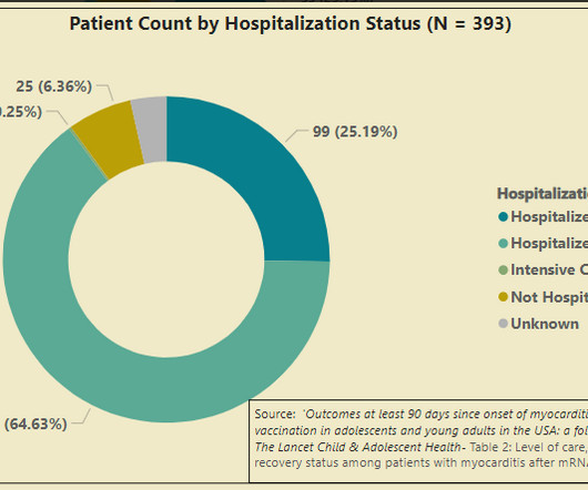
In summary, we have a CDC follow-up study that shows 25% of survey responders with vaccine myocarditis were admitted to the ICU, and one of these cases required a modified type of heart/lung bypass machine to stay alive. But a good long-term prognosis related to these cardiac scars is what everyone hopes for, not what anyone knows.
We organize all of the trending information in your field so you don't have to. Join thousands of users and stay up to date on the latest articles your peers are reading.
You know about us, now we want to get to know you!
Let's personalize your content
Let's get even more personalized
We recognize your account from another site in our network, please click 'Send Email' below to continue with verifying your account and setting a password.


Let's personalize your content