This site uses cookies to improve your experience. To help us insure we adhere to various privacy regulations, please select your country/region of residence. If you do not select a country, we will assume you are from the United States. Select your Cookie Settings or view our Privacy Policy and Terms of Use.
Cookie Settings
Cookies and similar technologies are used on this website for proper function of the website, for tracking performance analytics and for marketing purposes. We and some of our third-party providers may use cookie data for various purposes. Please review the cookie settings below and choose your preference.
Used for the proper function of the website
Used for monitoring website traffic and interactions
Cookie Settings
Cookies and similar technologies are used on this website for proper function of the website, for tracking performance analytics and for marketing purposes. We and some of our third-party providers may use cookie data for various purposes. Please review the cookie settings below and choose your preference.
Strictly Necessary: Used for the proper function of the website
Performance/Analytics: Used for monitoring website traffic and interactions
AI can make echocardiogram analysis easier for patients to understand, according to a study published July 31 in the Journal of the American College of Cardiology: Cardiovascular Imaging.
All underwent standard comprehensive echocardiography on a 5G cellular network robotic arm-based remote echocardiographic system, as well as a conventional echocardiographic platform (at Zhongshan Hospital) successively. tim.hodson Thu, 08/29/2024 - 11:39 Aug. 30 – Sept. If you enjoy this content, please share it with a colleague
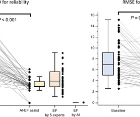
Methods This prospective, multicentre echocardiographic study was conducted by five cardiologists of level 1 echocardiographic skill (minimum level of competency to interpret images) from different hospitals. Protocol 1: Visual LVEFs for the 48 cases were measured without input from the AI-LVEF. ±3.2 (p=0.004) p=0.004) with AI-LVEF.
"Discover the latest guidelines from the European Society of Cardiology for managing chronic coronary syndromes, including the strong recommendation for using u
Echocardiogram was performed for patients with ECMO, including at pre-ECMO, during cannulation, during ECMO support, during the ECMO wean, and a follow up within 3 months after weaning.
Ascension Saint Thomas Hospital Improves Performance through Better Medical Record Management KCummings Fri, 03/29/2024 - 08:54 Overview In the fast-paced environment of healthcare, hospitals face significant challenges related to medical records. Accurate and relevant data from the STS Database uncovered areas for improvement.
The study will be launched at Cleveland Clinic and will focus on oHCM patients with the aim to validate the quality and accuracy of the monitoring echocardiograms performed by non-sonographers using UltraSights Real-Time Guidance software.
Our report describes two cases of SVS treated with endocardial ablation to improve LVOTO.Case reportCase 1: A 74-year-old female patient with angina and syncope was admitted to the hospital and diagnosed with SVS by transthoracic echocardiogram. After RFA was performed, the patient's symptoms significantly improved.
Smidt Heart Institute and Cedars-Sinai, both based in Los Angeles, used the largest dataset to date to trained a machine-learning algorithm that can interpret echocardiogram images.
Developed at Children’s National Hospital and detailed in the latest edition of the Journal of the American Heart Association , the new AI system combines the power of novel ultrasound probes with portable electronic devices installed with algorithms capable of diagnosing RHD on echocardiogram.
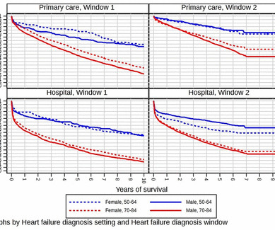
As a result, many heart failure cases go undiagnosed until symptoms force a specialist or emergency hospital visit, leading to worse patient outcomes and exacerbated healthcare costs.2 Survival and health economic outcomes in heart failure diagnosed at hospital admission versus community settings: a propensity-matched analysis.
After 24 hours, the patient was readmitted to the hospital with chest pain and troponin elevation, without ECG changes. A transthoracic echocardiogram (TTE) revealed a mobile mass on the right coronary cusp of the aortic valve ( figure 1 , ). The patient was discharged and apixaban was restarted 10 hours later.
We know that structural heart disease and heart failure are the leading causes of hospitalization and morbidity in the U.S. Data from the studies demonstrated that AISAP CARDIO enables non-cardiologist physicians to interpret point-of-care echocardiograms just as well as expert cardiologists of the MGB echocardiography lab.
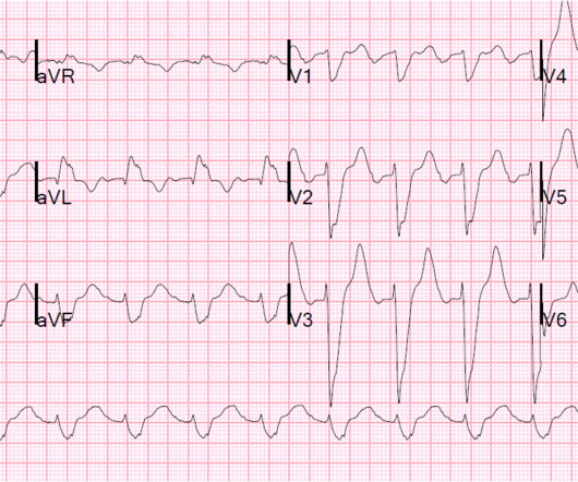
When flow is restored, wall motion may completely recover so that echocardiogram does not detect the previous ischemia. Transient ST elevation is very hazardous. Even when the serial troponins are negative, the ECG is critical to the diagnosis of ACS. This is not pericarditis because: a.
And then a slightly more remote past ECG Old inferior MI The patient's previous echocardiogram report was viewed: Decreased LV systolic performance, estimated left ventricular ejection fraction is 35%. Case continued The patient underwent an emergency formal echocardiogram and it was unchanged. Cath Lab activation was cancelled.
He was treated for infection and DKA and admission to hospital was planned. See this post: What do you think the echocardiogram shows in this case? Previously placed stents in the LAD (multiple) and mid circumflex and patent Formal echocardiogram: Normal left ventricular size and wall thickness.
He was admitted to our hospital with issues of chest pain, shortness of breath and heart palpitations without any obvious inducement. Figure 1 Transthoracic echocardiogram. (A) He had no medical history of myocardial infarction (MI). A 12-lead ECG indicated sinus rhythm with a heart rate of 78 bpm.
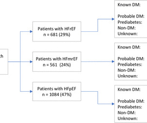
Patients’ HF phenotype was determined using the latest available echocardiogram. Results In total, 2326 patients (59% male, mean age 76±13 years) with HF and at least one echocardiogram were assessed. The number of patients with previous DM diagnosis was assessed.
He was intubated in the field and sedated upon arrival at the hospital. Echocardiogram showed LVEF 66% with normal wall motion and normal diastolic function. At his family's request, he was transferred to a hospital closer to his home to continue care. When EMS arrived the patient was in ventricular fibrillation.
David Austin, MD “We did not see evidence that we could reduce this biomarker of cardiotoxicity during chemotherapy [with an ACE inhibitor],” said David Austin, MD , consultant cardiologist at the academic cardiovascular unit in James Cook University Hospital , South Tees, Middlesbrough, United Kingdom, and the study’s lead author.
While in the hospital, he had witnessed ventricular fibrillation (VF) arrest for which he received external defibrillation. An echocardiogram showed newly reduced left ventricular ejection fraction of 30-35%.
The conventional TIA evaluation process often results in long ED stays or preventable hospital admissions. If indicated, the patient will be scheduled for an MRI within 24 hours and/or an echocardiogram within 48 hours of the TIA clinic visit. Radiology and cardiology have availability for urgent MRIs and echocardiograms.
Methods We identified all index hospitalisations with HF to John Hunter Hospital and Tamworth Rural Referral Hospital in the Hunter New England Local Health District over a 12 months. Results There were 664 patients who had an index HF admission to John Hunter and Tamworth hospitals in 2014. The median follow-up was 3.3
The patients were divided into dapagliflozin group and control group according to whether they took dapagliflozin during hospitalization. Two groups of patients' age, gender, diabetes duration, merge disease, echocardiogram and blood biochemical indexes, had no statistical difference (P>0.05).
He was admitted to the hospital for evaluation of these symptoms — but no ECG was done at that time. The rest of the patient’s hospital stay was uneventful and he was eventually discharged. An ECG was finally done at 9:17am on the 2nd hospital day. The consulting cardiologist wrote in their note: “Could be cardiac chest pain.
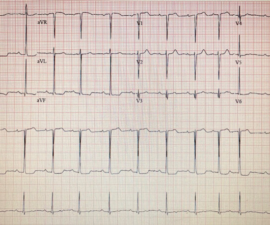
In this study of consecutive patients with LBBB who were hospitalized and had an echocardiogram, a QRS duration less than 170 ms (n = 262), vs. greater than 170 ms (n = 38), was associated with a significantly better ejection fraction (36% vs. 24%). Here is a similar case involving right bundle branch block.
Echocardiograms were performed with a standardised image acquisition protocol and reported by cardiologists. Results There were 465 individuals who underwent echocardiograms. Eligible persons aged 16–40 years were recruited according to a stratified randomised approach.
Methods:Data on healthcare providers and pharmacy certification, patient monitoring (from Patient Status Forms, based partly on echocardiograms), and screening for drug interactions prior to each dispense were collected.Results:Of 6,299 patients who received 1 dose of mavacamten, 60.0% were women; 64.6% were >60 years of age.
An echocardiogram showed severely reduced global systolic function with an EF of 20-25% and an LV apical thrombus. An echocardiogram showed an EF of 20-25%. The scan did not find PE, but showed evidence of coronary plaque: There are areas of dense white in the LAD (red and blue circles) and in the first diagonal (green circle).
for hospital-diagnosed patients). For the 9963 patients with symptoms recorded by their general practitioner before diagnosis, brain natriuretic peptide (BNP) testing was low, but echocardiogram use rose from 8.3% For those diagnosed in 2001/2002, the 5-year survival was 40.0% (40.2% vs 57.4%, compared with 33.9%
Objective To identify the most common transthoracic echocardiogram (TTE) parameters in patients hospitalised with severe acute respiratory syndrome coronavirus 2 (SARS-CoV-2/COVID-19) and their association with myocardial injury and outcomes. Results A total of 87 patients met the eligibility criteria. to 20.19, p<0.001).
Additionally, a bedside echocardiogram showed no wall motion abnormality and normal LV function. Patient 1 remained in the hospital overnight. A formal echocardiogram for patient 2 showed normal LV size, wall thickness, and global systolic function. Angiography revealed a 30% nonobstructive stenosis of the mid LAD.
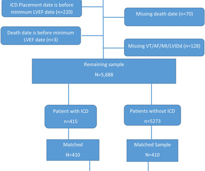
Methods Patients with lowest LVEF between 30% and 35% without an ICD prior to the lowest-LVEF echo (defined as ‘time zero’) were identified by querying echocardiography data from 28 November 2001 to 9 July 2020 at the Massachusetts General Hospital linked to ICD treatment status.
The purpose of this study was to determine the incidence of left and right ventricular dysfunction in patients hospitalized with acute COVID-19 and evaluate for cardiac recovery.Methods:A multicenter, retrospective cohort study was conducted. Adult patients were identified by hospitalizations using ICD-10 code U07.1
In the pre-hospital setting the varying modalities needed to rule-in/rule-out these causative factors are not available (eg, Chest X-ray, Echocardiogram, etc). Mechanical prosthetic valve Severe carotid artery stenosis is also implicated in embolic stroke.
Methods Patients from a large US hospital system undergoing combined LAAO and left-atrial CA from 8/2020 to 2/2024 were retrospectively analyzed and compared to a control group undergoing LAAO alone. vs. 19.4%, p =0.17) in-hospital complications were similar between the combined and control group, respectively.
This group was then further filtered to contain only those patients in whom the physician did NOT report severe aortic stenosis.Results:Between Jan and June 2023, 16,675 subjects ages 18-95 (avg 74) underwent transthoracic echocardiograms in our healthcare system, with 190 (1.1%) reported as having an AVA ≤ 1 cm2.
An echocardiogram showed: Left ventricular hypertrophy concentric. We found that 38% of out of hospital ventricular fibrillation was due to STEMI. Correlation of STEMI in Resuscitated Non-traumatic out-of-hospital Cardiopulmonary Arrest patients with Initial Rhythm and Cardiac Catheterization Findings (Abstract 580).
Transesophageal Echocardiogram (TEE) has traditionally been the gold standard for LAA clot detection. A total of 150 patients admitted to a tertiary hospital underwent both TTE and TEE before BMV, with TEE as the reference standard for evaluation.Results:The mean age of our study population was 41.4
Differences in outcomes frequencies were mostly confined to the primary MR subgroup, in which patients with above-mild MAC also experienced earlier, more frequent 2-year heart failure hospitalizations (20.8% METHODS:We retrospectively analyzed 968 individuals (median age, 79 [interquartile range, 70–86] years; 60.0% versus 9.6%;P=0.016;
Compared to their peers without the condition, people with atrial fibrillation are twice as likely to be admitted to hospital, five times more likely to have a stroke, three times more likely to develop heart failure, and twice as likely to die prematurely.4 Symptoms include palpitations, fatigue, shortness of breath, dizziness, and fainting.
Abstract Introduction Catheter ablation for atrial fibrillation (AF) reduces heart failure (HF) hospitalization in patients with HF with preserved ejection fraction (HFpEF). The primary endpoint was a composite of death from any cause or hospitalization for worsening HF.
On day 3 of hospitalization, he experienced a syncopal episode and had acute worsening of hypoxemia that prompted a CT angiography of the chest which revealed bilateral, large clot burden pulmonary emboli with proximal thrombus in both the right and left main pulmonary arteries. On arrival, his troponins were elevated 373, elevated BNP 934.
We organize all of the trending information in your field so you don't have to. Join thousands of users and stay up to date on the latest articles your peers are reading.
You know about us, now we want to get to know you!
Let's personalize your content
Let's get even more personalized
We recognize your account from another site in our network, please click 'Send Email' below to continue with verifying your account and setting a password.


Let's personalize your content