This site uses cookies to improve your experience. To help us insure we adhere to various privacy regulations, please select your country/region of residence. If you do not select a country, we will assume you are from the United States. Select your Cookie Settings or view our Privacy Policy and Terms of Use.
Cookie Settings
Cookies and similar technologies are used on this website for proper function of the website, for tracking performance analytics and for marketing purposes. We and some of our third-party providers may use cookie data for various purposes. Please review the cookie settings below and choose your preference.
Used for the proper function of the website
Used for monitoring website traffic and interactions
Cookie Settings
Cookies and similar technologies are used on this website for proper function of the website, for tracking performance analytics and for marketing purposes. We and some of our third-party providers may use cookie data for various purposes. Please review the cookie settings below and choose your preference.
Strictly Necessary: Used for the proper function of the website
Performance/Analytics: Used for monitoring website traffic and interactions
Before initiating therapeutic hypothermia, a head CT was done and showed fatal subarachnoid hemorrhage. However, she was found to have a fatal pontine hemorrhage and had a maximum troponin I, at 12 hours after presentation, of 2.0 Echocardiogram showed an anteroapical wall motion abnormality. This 81 yo was found comatose.
Herein, we present a case of recurrent left atrial myxoma with hemorrhagic, cerebral embolization.Case Report:A 34-year-old male presented with acute onset numbness and tingling of the left arm. Brain MRI demonstrated multiple hemorrhagic masses throughout the cerebral hemispheres with vasogenic edema.
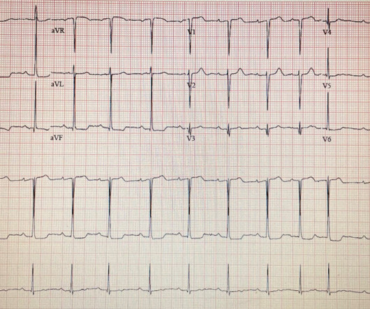
MRI Brain demonstrated Left MCA acute/subacute infarct, MCA/PCA watershed, and no hemorrhagic transformation. In the pre-hospital setting the varying modalities needed to rule-in/rule-out these causative factors are not available (eg, Chest X-ray, Echocardiogram, etc). No previous medical history was reported. We do have the ECG.
More often, tachycardia with ST segment abnormalities (elevation or depression) is due to an underlying illness (PE, sepsis, hemorrhage, dehydration, hypoxia, respiratory failure, etc.). Then ACS (STEMI) might be primary; this might be cardiogenic shock. One must clearly rule out these processes before jumping on the ACS diagnosis.
CTA head and neck were obtained and showed no evidence of intracranial hemorrhage, large vessel occlusion stroke (what a helpful and apt name for an acute arterial occlusion paradigm, by the way.), Echocardiogram was obtained and showed mild LVH without regional wall motion abnormality. Blood glucose was not low at 162 mg/dL.
Hopefully a repeat echocardiogram will be performed outpatient. Massive Transfusion for Motorcycle Collision with Hemorrhage, Troponin Elevated. Systolic function normal by visual assessment only, unable to visualize well for further characterization. 1900: RBBB and LAFB are almost fully resolved. 2300: QRS now within normal limits.
5) Myocardial contusion (edema and hemorrhage in the myocardium) which may result in dysrhythmias, blocks (especially RBBB as here), and poor cardiac contractility, including wall motion abnormalities. No further ECG, troponin, or echocardiogram was done because she was asymptomatic, and had a normal rhythm and rate.
See this case: what do you think the echocardiogram shows in this case? POCUS showed good LV-function and no pericardial effusion. Smith : It should be noted that, in subendocardial ischemia, in contrast to OMI, absence of wall motion abnormality is common. The patient had mild but diffuse abdominal tenderness.
An echocardiogram was done. A man in his 40s with multitrauma from motor vehicle collision Massive Transfusion for Motorcycle Collision with Hemorrhage, Troponin Elevated. Is there also Brugada? Here is the result: The estimated left ventricular ejection fraction is 50 %. There is no left ventricular wall motion abnormality identified.
I have ordered an echocardiogram which will be done today, after that patient can be discharged to home with follow-up in 2 to 3 months." The echo was normal. Learning points 1. In this regular wide complex tachycardia , since the rhythm converted w adenosine, it is almost certainly SVT w aberrancy, which can be either: A.
Due to the high risk of hemorrhagic conversion, the loading of antiplatelets was deferred. A 2D echocardiogram revealed an ejection fraction of 43%, hypokinesia of the anterior and intraventricular septum from base to apex, and severe mitral stenosis. ml subcutaneously once daily. An open-heart surgery was considered.
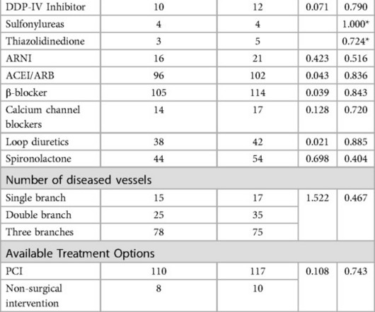
Two groups of patients' age, gender, diabetes duration, merge disease, echocardiogram and blood biochemical indexes, had no statistical difference (P>0.05). There were no significant differences in the number of coronary artery lesions, treatment regimens, cardiovascular and hypoglycemic drugs between the two groups (P>0.05).
In hemorrhagic strokes, there is a bleed and therefore the blood that would have gone to supply the brain cells goes somewhere else. It will help delineate between hemorrhagic and ischemic stroke. Another way of imaging the heart is via a transesophageal echocardiogram. This is important for several reasons.
Also consider non-hemorrhagic volume depletion, dehydration : orthostatic vitals may uncover this [see Mendu et al. (3)]. Serious outcomes included death, arrhythmia, myocardial infarction, structural heart disease, pulmonary embolism, and hemorrhage. Results : Presyncope constituted 0.5% of ED visits.
We organize all of the trending information in your field so you don't have to. Join thousands of users and stay up to date on the latest articles your peers are reading.
You know about us, now we want to get to know you!
Let's personalize your content
Let's get even more personalized
We recognize your account from another site in our network, please click 'Send Email' below to continue with verifying your account and setting a password.


Let's personalize your content