This site uses cookies to improve your experience. To help us insure we adhere to various privacy regulations, please select your country/region of residence. If you do not select a country, we will assume you are from the United States. Select your Cookie Settings or view our Privacy Policy and Terms of Use.
Cookie Settings
Cookies and similar technologies are used on this website for proper function of the website, for tracking performance analytics and for marketing purposes. We and some of our third-party providers may use cookie data for various purposes. Please review the cookie settings below and choose your preference.
Used for the proper function of the website
Used for monitoring website traffic and interactions
Cookie Settings
Cookies and similar technologies are used on this website for proper function of the website, for tracking performance analytics and for marketing purposes. We and some of our third-party providers may use cookie data for various purposes. Please review the cookie settings below and choose your preference.
Strictly Necessary: Used for the proper function of the website
Performance/Analytics: Used for monitoring website traffic and interactions
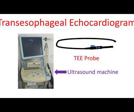
Echocardiogram is an image of the heart using ultrasound. The echo received from the body is processed by the computer in the machine to give an image of the heart. Usual echocardiogram is obtained by placing the transducer or probe on the chest. This is because the lungs cover part of the heart when you breath in.
In such scenarios, cardiologists’ primary form of treatment for children with coronary heart disease is a heartvalve implant. Unfortunately, the traditional pulmonary valves have a fixed diameter that can’t match the size of the child’s heart over time.
Usual colour Doppler echocardiogram is superimposition of colour Doppler images on a two dimensional echocardiogram. Colour M-Mode is superimposition of colour Doppler images on an M-Mode echocardiogram. All high velocity flows across the diseased valves will be shown as mosaic jets.
When the quantity is large enough to compress the heart, the person may feel breathless or dizzy because of a fall in blood pressure. Sometimes mild pericardial effusion may be detected by an echocardiogram done for other causes. Pericardial effusion is usually confirmed by an echocardiogram (ultrasound study of the heart).
So today i wanted to talk to you about what each heart test tells us about these different aspects of heart disease Tests that tell you about the heart as a pump The most commonly used test to assess the heart as a pump is an echocardiogram. If the heart has been left damaged, then that part of.
High Blood Pressure (Hypertension) Persistent high blood pressure forces the heart to work harder to pump blood. Over time, this additional strain causes the heart muscle to thicken, enlarging the heart. Antiarrhythmics to help manage abnormal heart rhythms. The following diagnostic tools are commonly used: 1.
These tests may include: Electrocardiogram (ECG) : Records the electrical activity of your heart. Echocardiogram : Uses sound waves to create images of your heart. Stress test : Assesses your heart’s function under stress. Blood tests : Measure cholesterol levels, blood sugar and other markers of heart health.
Common conditions that can cause our pump to become defective are: A previous heart attack – a heart attack means that a part of the heart has died and therefore, the pump has in some way become weaker Heartvalve disease – if our heartvalves are abnormally narrowed then they make it a lot more difficult for the heart to pump blood out.
This is to look for any mainly 4 things: To visualise a clot within the heart – which may sit in a small beak shaped structure of the atrium called atrial appendage or even in the ventricle. To look for any shunts within the heart. Another way of imaging the heart is via a transesophageal echocardiogram.
We organize all of the trending information in your field so you don't have to. Join thousands of users and stay up to date on the latest articles your peers are reading.
You know about us, now we want to get to know you!
Let's personalize your content
Let's get even more personalized
We recognize your account from another site in our network, please click 'Send Email' below to continue with verifying your account and setting a password.


Let's personalize your content