This site uses cookies to improve your experience. To help us insure we adhere to various privacy regulations, please select your country/region of residence. If you do not select a country, we will assume you are from the United States. Select your Cookie Settings or view our Privacy Policy and Terms of Use.
Cookie Settings
Cookies and similar technologies are used on this website for proper function of the website, for tracking performance analytics and for marketing purposes. We and some of our third-party providers may use cookie data for various purposes. Please review the cookie settings below and choose your preference.
Used for the proper function of the website
Used for monitoring website traffic and interactions
Cookie Settings
Cookies and similar technologies are used on this website for proper function of the website, for tracking performance analytics and for marketing purposes. We and some of our third-party providers may use cookie data for various purposes. Please review the cookie settings below and choose your preference.
Strictly Necessary: Used for the proper function of the website
Performance/Analytics: Used for monitoring website traffic and interactions
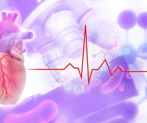
Getty Images milla1cf Tue, 01/16/2024 - 14:11 January 16, 2024 — Artificial intelligence (AI) has the potential to detect rheumatic heartdisease (RHD) with the same accuracy as a cardiologist, according to new research demonstrating how sophisticated deep learning technology can be applied to this disease of inequity.
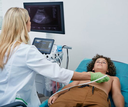
Food and Drug Adminstration (FDA) has approved DEFINITY (Perflutren Lipid Microsphere) as an ultrasound enhancing agent for use in pediatric patients with suboptimal echocardiograms, including those who have undergone heart transplant, or have Kawasaki disease or a congenital cardiovascular anomaly.
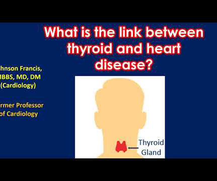
Heartdisease can occur with both increased function of the thyroid gland and decreased function of the thyroid gland. When thyroid function is increased, heart rate increases and the work load of the heart increases. In severe cases heart failure may occur.
Food and Drug Administration (FDA) has granted 510(k) clearance for its first-of-a-kind, AI-powered AISAP CARDIO point-of-care ultrasound (POCUS) software platform. We know that structural heartdisease and heart failure are the leading causes of hospitalization and morbidity in the U.S.
Introduction:Dextrocardia is a rare congenital condition where the heart's apex points to the right, with an incidence of about 0.01%. Patients usually have a normal life expectancy unless other structural heartdiseases are present. An intravascular ultrasound was also performed, which was negative for vessel dissection.
For example, by integrating Ventripoint’s AI-powered heart-scanning technology, which turns ultrasound images of the heart into MRI-quality heart images, InView provides pediatric cardiologists with access to MRI-quality heart images at a fraction of the cost and time needed for traditional MRIs.
So today i wanted to talk to you about what each heart test tells us about these different aspects of heartdisease Tests that tell you about the heart as a pump The most commonly used test to assess the heart as a pump is an echocardiogram. If the heart has been left damaged, then that part of.
During echocardiography, a transducer transmits the ultrasound beam towards the heart. Echoes received by the transducer from various structures of the heart are analysed by the echocardiograph and a graphical representation displayed on the monitor. The image shown here is an animated 2 dimensional echocardiogram.
It is also very important to mention a history of high blood pressure, diabetes, elevated cholesterol, family history of premature heartdisease, stroke or even sudden death. Ultrasound – this is easily available, very portable and usually a very low risk investigation. There are a variety of ways to look at these.
At first glance, the subject of heartdisease can seem exceptionally complex – consisting of several different conditions, medical jargon and very scary sounding terminology. As the heart becomes more muscular, it becomes stiffer and therefore does not fill with as much blood and therefore pumps less blood out.
Smith comment: This patient did not have a bedside ultrasound. Had one been done, it would have shown a feature that is apparent on this ultrasound (however, this patient's LV function would not be as good as in this clip): This is recorded with the LV on the right. In fact, bedside ultrasound might even find severe aortic stenosis.
His ED cardiac ultrasound (which is not at all ideal for detecting wall motion abnormalities, and is also very operator dependent for this finding) was significant for depressed global EF. I think a good start would be a posterior EKG and a high quality contrast echocardiogram read by an expert. What would you do in this scenario?
Check : [vitals, SOB, Chest Pain, Ultrasound] If the patient has Abdominal Pain, Chest Pain, Dyspnea or Hypoxemia, Headache, Hypotension , then these should be considered the primary chief complaint (not syncope). heart auscultation (aortic stenosis); c. h/o heartdisease (+1) 3. orthostatic vitals b.
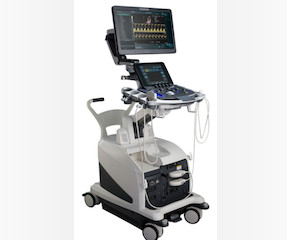
ai have partnered to equip Fujifilms LISENDO 800 cardiovascular ultrasound system with Us2.ai's ais software, when used with the LISENDO 880 ultrasound system, fully automates the analysis and reporting of echocardiograms and provides comprehensive cardiac measurements for the diagnosis of heartdisease.
We organize all of the trending information in your field so you don't have to. Join thousands of users and stay up to date on the latest articles your peers are reading.
You know about us, now we want to get to know you!
Let's personalize your content
Let's get even more personalized
We recognize your account from another site in our network, please click 'Send Email' below to continue with verifying your account and setting a password.


Let's personalize your content