This site uses cookies to improve your experience. To help us insure we adhere to various privacy regulations, please select your country/region of residence. If you do not select a country, we will assume you are from the United States. Select your Cookie Settings or view our Privacy Policy and Terms of Use.
Cookie Settings
Cookies and similar technologies are used on this website for proper function of the website, for tracking performance analytics and for marketing purposes. We and some of our third-party providers may use cookie data for various purposes. Please review the cookie settings below and choose your preference.
Used for the proper function of the website
Used for monitoring website traffic and interactions
Cookie Settings
Cookies and similar technologies are used on this website for proper function of the website, for tracking performance analytics and for marketing purposes. We and some of our third-party providers may use cookie data for various purposes. Please review the cookie settings below and choose your preference.
Strictly Necessary: Used for the proper function of the website
Performance/Analytics: Used for monitoring website traffic and interactions
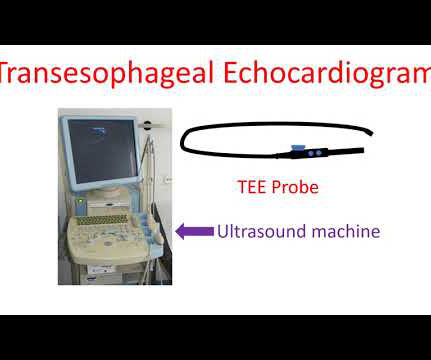
Echocardiogram is an image of the heart using ultrasound. An ultrasound beam is transmitted into the body using a device known as transducer. Transesophageal echocardiogram or TEE test, is obtained by introducing a special type of transducer, also called a TEE probe, through the throat into the food pipe (esophagus) and stomach.
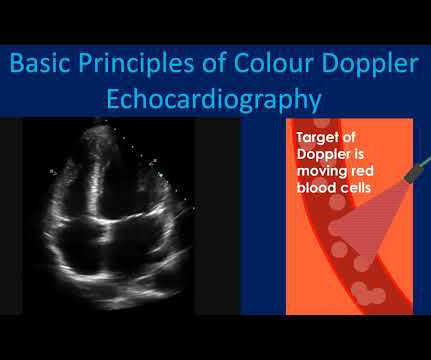
Usual colour Doppler echocardiogram is superimposition of colour Doppler images on a two dimensional echocardiogram. Colour M-Mode is superimposition of colour Doppler images on an M-Mode echocardiogram. Colour Doppler echocardiography receives the ultrasound signals reflected from moving red blood cells in the heart.
Ultrasound image of the heart – echocardiogram, showing fluid collection around the heart, marked as PE, short for pericardial effusion. Collection of fluid within the covering of the heart is called pericardial effusion. If it is severe enough to compress the heart, it prevents proper filling of the heart and blood pressure falls.
Sometimes mild pericardial effusion may be detected by an echocardiogram done for other causes. Pericardial effusion is usually confirmed by an echocardiogram (ultrasound study of the heart). When the quantity is large enough to compress the heart, the person may feel breathless or dizzy because of a fall in blood pressure.
During echocardiography, a transducer transmits the ultrasound beam towards the heart. The image shown here is an animated 2 dimensional echocardiogram. This one is an older mode known as time-motion mode or M-Mode echocardiogram. Hence a basic knowledge is needed for all physicians and paramedics.
We organize all of the trending information in your field so you don't have to. Join thousands of users and stay up to date on the latest articles your peers are reading.
You know about us, now we want to get to know you!
Let's personalize your content
Let's get even more personalized
We recognize your account from another site in our network, please click 'Send Email' below to continue with verifying your account and setting a password.


Let's personalize your content