This site uses cookies to improve your experience. To help us insure we adhere to various privacy regulations, please select your country/region of residence. If you do not select a country, we will assume you are from the United States. Select your Cookie Settings or view our Privacy Policy and Terms of Use.
Cookie Settings
Cookies and similar technologies are used on this website for proper function of the website, for tracking performance analytics and for marketing purposes. We and some of our third-party providers may use cookie data for various purposes. Please review the cookie settings below and choose your preference.
Used for the proper function of the website
Used for monitoring website traffic and interactions
Cookie Settings
Cookies and similar technologies are used on this website for proper function of the website, for tracking performance analytics and for marketing purposes. We and some of our third-party providers may use cookie data for various purposes. Please review the cookie settings below and choose your preference.
Strictly Necessary: Used for the proper function of the website
Performance/Analytics: Used for monitoring website traffic and interactions
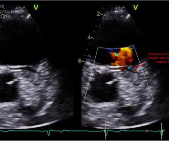
The patient was referred for an exercise nuclear study and did 11 min on the Bruce protocol without angina or ischaemic ECG changes. A transthoracic echocardiogram was performed ( figure 1 ). Figure 1 Transthoracic echocardiogram ((A) apical four-chamber view; (B) parasternal short-axis view).
Take walks, dance to holiday music, or engage in short bursts of exercise throughout the day. Regular check-ups allow your cardiologist to detect these issues through tests like blood work, EKGs, and echocardiograms. Focus on balanced meals with plenty of fruits, vegetables, and lean protein.
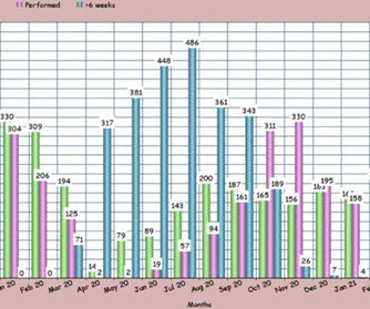
Objective To assess the feasibility, efficacy and safety of performing exercise stress echocardiography (ESE) for the assessment of myocardial ischaemia during the COVID-19 pandemic. Propensity-matched data showed no significant difference in exercise workload between patients undergoing ESE during and prepandemic.
While much attention is given to modifiable risk factors such as diet, exercise, and smoking, the role of genetics in heart disease is equally critical yet less understood by the general public. Exercise: Regular physical activity strengthens the heart and improves blood circulation. Heart imaging, such as echocardiograms or CT scans.
Objective To evaluate for correlation between exercise capacity as assessed by peak oxygen consumption (pVO 2 ) measurement during a cardiopulmonary exercise test (CPET) and smartwatches reporting this parameter in patients with adult congenital heart disease (ACHD) complex lesions.
EECP can improve exercise tolerance, reduce anginal symptoms, and enhance endothelial function, offering a potential alternative for patients with ischemic HFrEF awaiting heart transplantation.Description of a Case:A 58-year-old Hispanic female with a history of severe CAD, type II DM, hyperlipidemia, and CVA.
The echocardiogram showed normal cardiac structure and function, however, there was a concern for possible anomalous origin of the left coronary artery. Treadmill exercise stress test showed excellent functional capacity without exercise-induced chest pain or ischemic ECG changes.
Echocardiogram An echocardiogram uses sound waves to produce a detailed image of the heart, allowing doctors to see the size of the heart chambers and how well the heart is pumping blood. Exercise regularly to keep the heart strong and healthy. The following diagnostic tools are commonly used: 1.
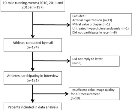
Methods Healthy, normotensive, male Caucasian participants of a 10-mile race were assessed with a 2D echocardiogram and comprehensive interview. Results Ninety-two of 121 athletes (aged 42±8 years) had sufficient echocardiogram quality and were used for analysis. Aortic strain, AD and aortic stiffness index were calculated.
After 5 weeks of the diet, HFD mice and their littermate controls underwent baseline exercise testing and echocardiograms. Echocardiograms were performed under 1.5% inhalational isoflurane.
Yes, COVID-19 symptoms can resemble a heart attack, including chest pain, shortness of breath, and changes in echocardiogram or EKG. Echocardiogram: Utilizing sound waves, this test generates images of the heart and its chambers to identify potential issues. Can COVID-19 symptoms mimic a heart attack?
Background:Cardiac output reserve and exercise capacity are strong predictors of life expectancy. Chronotropic incompetence (CI) is the inability to reach an age appropriate maximum heart rate with exercise. CI reduces cardiac output reserve and exercise capacity, both of which increase all-cause mortality risk. x age in years).Results:The
Echocardiogram : Uses sound waves to create images of your heart. Heart disease prevention : By identifying risk factors for heart disease such as smoking, unhealthy diet and lack of exercise you can take steps to modify your lifestyle and reduce your risk. Stress test : Assesses your heart’s function under stress.
Sent by anonymous A man in his 40s with no previous heart disease presented within 30 minutes of onset of acute chest pain that started while exercising. Formal echocardiogram: Systolic function is at the lower limits of normal. Written by Pendell Meyers with edits by Smith. Some function might possibly recover over weeks.
She underwent exerciseechocardiogram in mid October where she exercised for nearly 7 minutes on the standard Bruce protocol and had typical anginal pain and shortness of breath. Baseline echocardiogram showed moderate LV systolic dysfunction with no wall motion abnormalities. There is inferoseptal hypokinesis.
On this visit, he expressed worsening exercise tolerance, new orthopnea, and he told his provider that the omeprazole did not relieve any symptoms. An echocardiogram showed severely reduced global systolic function with an EF of 20-25% and an LV apical thrombus. An echocardiogram showed an EF of 20-25%.
During aerobic exercise which is isotonic, the heart rate and stroke volume increases. Isometric exercise or weight training on the other hand causes only slight increase in cardiac output due to increase in heart rate. Effect of exercise on right ventricle. J point elevation and early repolarization pattern has been reported.
But, still for an academic exercise, we will try. Similarly, for echocardiogram, what we would do usually is, first we do a clinical history evaluation, then physical examination, and after that only we proceed with echocardiography in our routine work. This is an echocardiographic image from the parasternal long axis view.
So today i wanted to talk to you about what each heart test tells us about these different aspects of heart disease Tests that tell you about the heart as a pump The most commonly used test to assess the heart as a pump is an echocardiogram. Overall though a normal cardiac MRI is even more reassuring than a normal echocardiogram.
The problem with both these tests are that they study the heart at rest and sometimes abnormalities may be picked up only when the heart is stressed and therefore combining these modalities with a stress test can be even more helpful and by far the best form of stress is exercise.
He visited an outpatient clinic for it and an echocardiogram and exercise stress test was normal. In the meantime, cardiology consultant sees the patient and performs a bedside echocardiogram which revealed no major wall motion abnormalities. He has 40 packs-year of smoking history. He denies taking any medication.
This integration enables cardiologists to access and review imaging studies directly within the EHR platform, such as echocardiograms, stress tests, and angiograms. Moreover, patients can receive personalized reminders, track their exercise routines, and monitor vital signs using compatible wearable devices.
Echocardiogram showing thickened interventricular septum and mitral regurgitation in HCM. Dynamic gradient may be sought by glyceryl trinitrate, Valsalva manoeuvre, standing position or even symptom limited exercise. Obstruction may occur during recovery and post exercise monitoring of gradient is mandatory.
I have ordered an echocardiogram which will be done today, after that patient can be discharged to home with follow-up in 2 to 3 months." The echo was normal. Learning points 1. In this regular wide complex tachycardia , since the rhythm converted w adenosine, it is almost certainly SVT w aberrancy, which can be either: A.
As an exercise, lets calculate the equation for differentiating the ST elevation between benign early repolarization and LAD occlusion. No further echocardiograms were available after cath. LAD occlusion. Great case. Only viewed on my phone." The patient was discharged one day after intervention and appears to be doing well.
Echocardiogram: Estimated left ventricular ejection fraction, lower limits of normal; 45-50%. In my experience — sinus tach rarely exceeds 170/minute in a non-exercising adult patient. Tele Monitor: Normal sinus rhythm throughout, no ectopic atrial or ventricular beats. Regional wall motion abnormality-inferior/inferoseptum: akinetic.
Next day, a stress echo was done: The exercise stress echocardiogram is normal. Normal estimated left ventricular ejection fraction improved with stress. No wall motion abnormality at rest. No wall motion abnormality with stress. The stress electrocardiogram is non-diagnostic. The patient did not report angina with stress.
Previously healthy, taking no medication and exercising regularly. No anginal symptoms asymptomatic during physical exercise. It is reasonable to perform an echocardiogram to evaluate LV function. Below in Figure-1 is this patient's admission ECG. How will you manage this patient? or is wider than 130 msec.,
Transthoracic echocardiogram, bilateral carotid Doppler ultrasound, and electrocardiogram were normal. A treadmill exercise test revealed ischemic changes. No previous history of hypertension or diabetes. There was no abnormality in physical examination.
We organize all of the trending information in your field so you don't have to. Join thousands of users and stay up to date on the latest articles your peers are reading.
You know about us, now we want to get to know you!
Let's personalize your content
Let's get even more personalized
We recognize your account from another site in our network, please click 'Send Email' below to continue with verifying your account and setting a password.


Let's personalize your content