This site uses cookies to improve your experience. To help us insure we adhere to various privacy regulations, please select your country/region of residence. If you do not select a country, we will assume you are from the United States. Select your Cookie Settings or view our Privacy Policy and Terms of Use.
Cookie Settings
Cookies and similar technologies are used on this website for proper function of the website, for tracking performance analytics and for marketing purposes. We and some of our third-party providers may use cookie data for various purposes. Please review the cookie settings below and choose your preference.
Used for the proper function of the website
Used for monitoring website traffic and interactions
Cookie Settings
Cookies and similar technologies are used on this website for proper function of the website, for tracking performance analytics and for marketing purposes. We and some of our third-party providers may use cookie data for various purposes. Please review the cookie settings below and choose your preference.
Strictly Necessary: Used for the proper function of the website
Performance/Analytics: Used for monitoring website traffic and interactions
Patients undergoing atrial fibrillation (AFib) ablation who were not properly anticoagulated and did not undergo preprocedural transesophageal echocardiogram (TEE) were significantly more likely to suffer from transient ischemic attack (TIA) or pulmonary embolism (PE).
Patients with lead-associated vegetation larger than 10 mm face increased odds of embolism and mortality. Percutaneous mechanical aspiration devices reduce the risk of embolization during lead extraction. Cardiac implantable electronic device(CIED) extraction is recommended in patients with related infective endocarditis (IE).
Background and Purpose:Right-to-left shunt (RLS) is one of the potential embolic sources in embolic stroke of undetermined source (ESUS), but the eligibility of conducting shunt study to detect RLS in ESUS is still unknown. Stroke, Volume 55, Issue Suppl_1 , Page ATP259-ATP259, February 1, 2024.
Introduction:Atrial myxomas are cardiac tumors that can cause arterio-occlusive diseases due to embolization of myxomatous fragments. Repeat echocardiograms did not show recurrence of a myxoma. Circulation, Volume 150, Issue Suppl_1 , Page A4140574-A4140574, November 12, 2024.
CT of the chest showed no pulmonary embolism but bibasilar infiltrates. EKG with paced complexes shown below shows much narrower QRS complex and echocardiogram showed improved LV systolic function primarily due to improvement in LV dyssynchrony. (J She was intubated. Bedside cardiac ultrasound showed moderately decreased LV function.
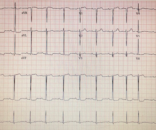
Examples of cardio embolic stroke etiology include: 1. Atrial Fibrillation 2. Cardiomyopathy with mural thrombus 3. Patent Foramen Ovale 4. Severe calcific Aortic (valve) Stenosis 5. Mechanical prosthetic valve Severe carotid artery stenosis is also implicated in embolic stroke. Here is the admission ECG.
Transthoracic echocardiogram (TTE) showed an ejection fraction (EF) of 40% and a moderate-large pericardial effusion with signs of tamponade. Intra-operative transesophageal echocardiogram (TEE) post-decannulation showed a normal EF without segmental abnormalities. Electrocardiogram (EKG) was unremarkable.
Transthoracic echocardiogram revealed right to left shunting consistent with a patent foramen ovale (PFO). She was initiated on thrombolytic therapy and experienced resolution of symptoms.
A transthoracic echocardiogram (TTE) was ordered and showed a linear highly mobile mass in the right atrium that was intermittently protruding into the right ventricle and into the main pulmonary artery through the pulmonic valve. There was no visible evidence of tumor extension of the renal mass into the inferior vena cava.
BackgroundComplex aortic plaque (CAP) is a potential embolic source in patients with cryptogenic stroke (CS). Journal of the American Heart Association, Volume 12, Issue 23 , December 5, 2023.
The commonest causes of MINOCA include: atherosclerotic causes such as plaque rupture or erosion with spontaneous thrombolysis, and non-atherosclerotic causes such as coronary vasospasm (sometimes called variant angina or Prinzmetal's angina), coronary embolism or thrombosis, possibly microvascular dysfunction.
Echocardiogram showing thickened interventricular septum and mitral regurgitation in HCM. Mid cavity obstruction in HCM is associated with apical aneurysm, systemic embolism, and arrhythmias. SAM in HCM Systolic anterior movement of mitral valve occurs in 30 – 60%, but it is not specific. Doppler echo showing LVOT gradient in HCM.
The next morning the patient went for his routine echocardiogram, where the operator noticed a dilated aortic root at 5.47 Patients with pulmonary embolism or aortic dissection who have normal variant ST elevation are at high risk of being diagnosed with pericarditis when what they have is far more serious!!
Here is the cath report: Echocardiogram: There is severe hypokinesis of entire LV apex and apical segment of all the walls. Transient and partial thrombosis at the site of a non-obstructive plaque with subsequent spontaneous fibrinolysis and distal embolization may be one of the mechanisms responsible for the occurrence of MINOCA.
Echocardiogram An echocardiogram uses sound waves to produce a detailed image of the heart, allowing doctors to see the size of the heart chambers and how well the heart is pumping blood. Blood Clots: An enlarged heart is more prone to developing blood clots, which can lead to stroke or pulmonary embolism.
He was started on a heparin drip and CTA of the chest was ordered to rule out pulmonary embolism. Echocardiogram showed severe RV dilation with McConnell’s sign and an elevated RVSP. Electrocardiographic Differentiation Between Acute Pulmonary Embolism and Acute Coronary Syndromes on the Basis of Negative T Waves - ScienceDirect.
Despite his large clot burden, there was absence of obstructive shock.Transthoracic Echocardiogram and bilateral duplex venous ultrasound were obtained to evaluate for right heart strain and clot burden. this clinical case, we discussed sub-massive pulmonary embolism (PE) complicated by a right heart clot-in-transit presenting insidiously.
He visited an outpatient clinic for it and an echocardiogram and exercise stress test was normal. Bi-phasic scan showed no dissection or pulmonary embolism. In the meantime, cardiology consultant sees the patient and performs a bedside echocardiogram which revealed no major wall motion abnormalities.
While awaiting transfer to the cath lab, STAT echocardiogram was performed and showed LVEF 30-35%, as well as anterior, inferior, and apical hypokinesis, and apical thrombus. Then, part of the thrombus embolized into the LCx causing an inferoposterolateral OMI. (As This confirms the suspicion of prior anterior OMI.
Cardiac enzymes, CTs, echocardiograms, carotid ultrasounds, and electroencephalography all affected diagnosis or management in Postural blood pressure , performed in only 38% of episodes, had the highest yield with respect to affecting diagnosis (18-26%) or management (25-30%) and determining etiology of the syncopal episode (15-21%).
Case continued Troponins over 26 hours, from right to left : Echocardiogram: Mild concentric left ventricular wall thickening, normal cavity size, and normal systolic function. By itself seeing this ECG pattern does not necessarily mean that the patient has a pulmonary embolism. The estimated left ventricular ejection fraction is 64%.
Despite apparently hearing the above history together with two diagnostic ECGs and a troponin compatible with OMI, the cardiologist thought the ECG represented pericarditis and recommended echocardiogram. Echocardiogram was finally performed five hours after the first diagnostic ECG. This far out, the benefit of PCI is very attenuated.
We organize all of the trending information in your field so you don't have to. Join thousands of users and stay up to date on the latest articles your peers are reading.
You know about us, now we want to get to know you!
Let's personalize your content
Let's get even more personalized
We recognize your account from another site in our network, please click 'Send Email' below to continue with verifying your account and setting a password.


Let's personalize your content