This site uses cookies to improve your experience. To help us insure we adhere to various privacy regulations, please select your country/region of residence. If you do not select a country, we will assume you are from the United States. Select your Cookie Settings or view our Privacy Policy and Terms of Use.
Cookie Settings
Cookies and similar technologies are used on this website for proper function of the website, for tracking performance analytics and for marketing purposes. We and some of our third-party providers may use cookie data for various purposes. Please review the cookie settings below and choose your preference.
Used for the proper function of the website
Used for monitoring website traffic and interactions
Cookie Settings
Cookies and similar technologies are used on this website for proper function of the website, for tracking performance analytics and for marketing purposes. We and some of our third-party providers may use cookie data for various purposes. Please review the cookie settings below and choose your preference.
Strictly Necessary: Used for the proper function of the website
Performance/Analytics: Used for monitoring website traffic and interactions
By applying AI to echocardiograms, we can help clinicians more easily detect the signs of heart valve disease so that patients get the care they need as soon as possible. Investigators trained a deep-learning program to flag patterns of tricuspid regurgitation in 47,312 echocardiograms done at Cedars-Sinai between 2011 and 2021.
These patients offer unique inherent and surgically altered anatomic challenges for electrophysiology procedures. Adults with congenital heart disease (ACHD) are surviving to increasingly older ages.
Transthoracic echocardiogram revealed normal biventricular function and dimension. Holter monitor showed 28% burden of PVCs with various morphologies consistent with right ventricular (RV) inflow and outflow tract exits.
Methods We conducted an analysis of all patients who had received either a single or dual lead cardiac implantable electronic devices, excluding biventricular devices, and had a prior transthoracic echocardiogram demonstrating an ejection fraction of less than 50%.
ai’s echocardiogram algorithms, which automate measurements and pre-populate structured report templates, InView eliminates manual steps and improving the speed of coordination-of-care for patients with suspected heart disease. As well, by incorporating Us2.ai’s
vs. 4.5%, p =0.96) on transesophageal echocardiogram did not differ. Both major (1.4% vs. 2.1%, p =0.72) and minor (27.8% vs. 19.4%, p =0.17) in-hospital complications were similar between the combined and control group, respectively. At 45 days, presence of peri-device leak (18.3% vs. 30.4%, p =0.07) and device related thrombosis (4.5%
Afterward, a transesophageal echocardiogram guided implantation of both a Micra AV 2 (Medtronic) leadless pacemaker in the interventricular septum within the right ventricle and an Aveir (Abbott) leadless pacemaker in the superior base of the right atrial appendage was performed with successful pacing.
Jesse McLaren @ECGcases [link] [link] This case was kindly submitted by Dr. Paco Dardon (@PacoDardon), and it’s a privilege to present it as a formal review due to the many pathophysiological, and electrophysiological, phenomenon at play. Indeed, bedside Echocardiogram revealed severe left ventricular impairment of Takotsubo cardiomyopathy.
Patients who received pacemakers for an advanced atrioventricular block or bradycardia with atrial fibrillation, baseline LV ejection fraction (LVEF) ≥ 50%, and echocardiogram recorded at least 6 months postimplantation were included. The paced QRS recorded immediately after implantation was analyzed.
After excluding patients with congenital or rheumatic heart disease, heart transplant recipients, or those without baseline echocardiogram, a total of 130 patients were included in the analysis. Echocardiographic data were analyzed at baseline before ablation, and at early follow-up within 1-year postablation.
Cupid EHR from Epic boasts the following: Cloud-based EHR Offers integrated order entry, scheduling, procedure documentation, structured reporting, and data analytics for cardiology practices Supports a wide range of workflows, including Echocardiograms, Ultrasound vascular, Cardiac Cath, stress testing, Electrophysiology, and structured documentation (..)
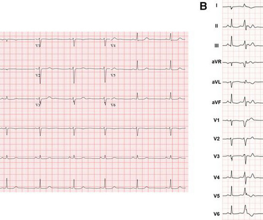
An echocardiogram was done. 2 weeks Here is the final electrophysiology note: It is unclear what precipitated his motor vehicle collision. Is there also Brugada? Here is the result: The estimated left ventricular ejection fraction is 50 %. There is no left ventricular wall motion abnormality identified. Right ventricular prominence.
I have ordered an echocardiogram which will be done today, after that patient can be discharged to home with follow-up in 2 to 3 months." Admission and referral to electrophysiology is always indicated. The echo was normal. Learning points 1. These tachydysrhythmias are so fast that they can degenerate into ventricular fibrillation.
It is also published in Heart Rhythm , the official journal of the HRS, Journal of Arrhythmia , the official journal of the APHRS, and Journal of Interventional Cardiac Electrophysiology , the official journal of the LAHRS. 7 Atrial fibrillation has a significant impact on people’s lives.
He had his echocardiogram done already and was normal. Clinical and electrophysiologic findings in patients with paroxysmal slowing of the sinus rate and apparent Mobitz type II atrioventricular block. Pacing and Clinical Electrophysiology, 35(7), e210–e213. I didn’t have calipers to measure the PR accurately though.
HFpEF was diagnosed from a history of congestive HF and/or combined criteria of N-terminal pro-brain natriuretic peptide (NT-proBNP) concentration and transthoracic echocardiogram parameters, including average septal-lateral E/e' and tricuspid regurgitation peak velocity.
Formal echocardiogram showed normal EF, no wall motion abnormalities, no pericardial effusion. She has not yet been seen by electrophysiology or had further genetic testing for Brugada syndrome. The patient proceeded to cath where all coronaries were described as normal with no evidence of any CAD, spasm, or any other abnormality.
A formal echocardiogram was completed the next day and again showed a normal ejection fraction without any focal wall motion abnormalities to suggest CAD. She has not yet been seen by electrophysiology or had further genetic testing for Brugada syndrome. The Troponin I was cycled over time and was 0.353 followed by 0.296.
Later, he underwent a formal echocardiogram: Very severe left ventricular enlargement (LVED diameter 7.4 Patient course The patient was started on beta blockers and schedule for an electrophysiologic study. A bedside POC cardiac ultrasound was done: Findings: Decreased left ventricular systolic function. Try adenosine.
Cardiac enzymes, CTs, echocardiograms, carotid ultrasounds, and electroencephalography all affected diagnosis or management in Postural blood pressure , performed in only 38% of episodes, had the highest yield with respect to affecting diagnosis (18-26%) or management (25-30%) and determining etiology of the syncopal episode (15-21%).
We organize all of the trending information in your field so you don't have to. Join thousands of users and stay up to date on the latest articles your peers are reading.
You know about us, now we want to get to know you!
Let's personalize your content
Let's get even more personalized
We recognize your account from another site in our network, please click 'Send Email' below to continue with verifying your account and setting a password.


Let's personalize your content