Remove Echocardiogram Remove Electrocardiogram Remove Laboratory
article thumbnail

Case Report: Lacosamide unmasking SCN5A-associated Brugada syndrome in a young female with epilepsy

Frontiers in Cardiovascular Medicine

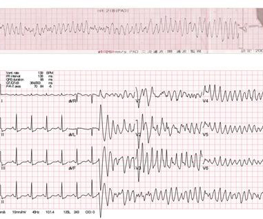
Workup including routine laboratory results, 12-lead electrocardiogram (ECG), echocardiogram, and coronary angiogram was non-specific. During the intravenous lacosamide infusion, the patient developed sudden cardiac arrest caused by ventricular arrhythmias necessitating resuscitation.

article thumbnail

Interplay between mitochondrial dysfunction and lysosomal storage: challenges in genetic metabolic muscle diseases with a focus on infantile onset Pompe disease

Frontiers in Cardiovascular Medicine

This comprehensive evaluation included the use of ultrasound echocardiograms, computed tomography (CT) scans, electrocardiograms, mutagenesis analysis, and structural analysis to gain insights into the patient's condition and the underlying mechanisms of PD.

article thumbnail

Emergency Department Syncope Workup: After H and P, ECG is the Only Test Required for Every Patient.

Dr. Smith's ECG Blog

Abnormal Electrocardiogram (ECG): Defined (San Fran syncope rule) as any new changes when compared to the last ECG or presence of non-sinus rhythm. Results : Electrocardiograms (99%), telemetry (95%), cardiac enzymes (95%), and head computed tomography (CT) (63%) were the most frequently obtained tests.