This site uses cookies to improve your experience. To help us insure we adhere to various privacy regulations, please select your country/region of residence. If you do not select a country, we will assume you are from the United States. Select your Cookie Settings or view our Privacy Policy and Terms of Use.
Cookie Settings
Cookies and similar technologies are used on this website for proper function of the website, for tracking performance analytics and for marketing purposes. We and some of our third-party providers may use cookie data for various purposes. Please review the cookie settings below and choose your preference.
Used for the proper function of the website
Used for monitoring website traffic and interactions
Cookie Settings
Cookies and similar technologies are used on this website for proper function of the website, for tracking performance analytics and for marketing purposes. We and some of our third-party providers may use cookie data for various purposes. Please review the cookie settings below and choose your preference.
Strictly Necessary: Used for the proper function of the website
Performance/Analytics: Used for monitoring website traffic and interactions
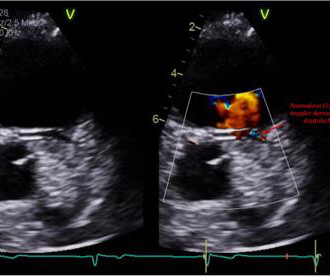
Electrocardiogram in clinic showed sinus arrhythmia with early repolarization and no ischemic changes. The echocardiogram showed normal cardiac structure and function, however, there was a concern for possible anomalous origin of the left coronary artery. Invasive coronary angiography ruled out luminal narrowing or dynamic compression.
Yes, COVID-19 symptoms can resemble a heart attack, including chest pain, shortness of breath, and changes in echocardiogram or EKG. Electrocardiograms (EKG or ECG): This diagnostic method effectively detects irregular heartbeats or arrhythmias by analyzing the electrical signals in the heart.
Sent by anonymous A man in his 40s with no previous heart disease presented within 30 minutes of onset of acute chest pain that started while exercising. Formal echocardiogram: Systolic function is at the lower limits of normal. Written by Pendell Meyers with edits by Smith. Some function might possibly recover over weeks.
The attending provider wrote “Agree with electrocardiogram interpretation”. On this visit, he expressed worsening exercise tolerance, new orthopnea, and he told his provider that the omeprazole did not relieve any symptoms. An echocardiogram showed an EF of 20-25%. The computer diagnostic algorithm diagnosed “Sinus rhythm.
Next day, a stress echo was done: The exercise stress echocardiogram is normal. The stress electrocardiogram is non-diagnostic. Normal estimated left ventricular ejection fraction improved with stress. No wall motion abnormality at rest. No wall motion abnormality with stress. The patient did not report angina with stress.
Echocardiogram An echocardiogram uses sound waves to produce a detailed image of the heart, allowing doctors to see the size of the heart chambers and how well the heart is pumping blood. Exercise regularly to keep the heart strong and healthy. Avoid excessive alcohol and drug use.
These tests may include: Electrocardiogram (ECG) : Records the electrical activity of your heart. Echocardiogram : Uses sound waves to create images of your heart. Exercise regularly : Aim for at least 30 minutes of moderate-intensity exercise most days. Stress test : Assesses your heart’s function under stress.
Smith , d and Muzaffer Değertekin a DIFOCCULT: DIagnostic accuracy oF electrocardiogram for acute coronary OCClUsion resuLTing in myocardial infarction. He visited an outpatient clinic for it and an echocardiogram and exercise stress test was normal. His first electrocardiogram ( ECG) is given below: --Sinus bradycardia.
Below are some of the most common causes of heart murmurs: Increased Blood Flow: Innocent heart murmurs often occur when there is an increase in blood flow, such as during pregnancy, exercise, fever, or growth spurts in children. In adults, innocent murmurs can develop in response to temporary factors like pregnancy, fever, or exercise.
Transthoracic echocardiogram, bilateral carotid Doppler ultrasound, and electrocardiogram were normal. A treadmill exercise test revealed ischemic changes. No previous history of hypertension or diabetes. There was no abnormality in physical examination.
We organize all of the trending information in your field so you don't have to. Join thousands of users and stay up to date on the latest articles your peers are reading.
You know about us, now we want to get to know you!
Let's personalize your content
Let's get even more personalized
We recognize your account from another site in our network, please click 'Send Email' below to continue with verifying your account and setting a password.


Let's personalize your content