This site uses cookies to improve your experience. To help us insure we adhere to various privacy regulations, please select your country/region of residence. If you do not select a country, we will assume you are from the United States. Select your Cookie Settings or view our Privacy Policy and Terms of Use.
Cookie Settings
Cookies and similar technologies are used on this website for proper function of the website, for tracking performance analytics and for marketing purposes. We and some of our third-party providers may use cookie data for various purposes. Please review the cookie settings below and choose your preference.
Used for the proper function of the website
Used for monitoring website traffic and interactions
Cookie Settings
Cookies and similar technologies are used on this website for proper function of the website, for tracking performance analytics and for marketing purposes. We and some of our third-party providers may use cookie data for various purposes. Please review the cookie settings below and choose your preference.
Strictly Necessary: Used for the proper function of the website
Performance/Analytics: Used for monitoring website traffic and interactions
Researchers used an AI-enabled digital stethoscope that captures electrocardiogram ( ECG ) data and heart sounds to identify twice as many cases of peripartum cardiomyopathy as compared to regular care, according to a news release from the American Heart Association.
A test phase included 33 patients who underwent transthoracic echocardiogram with ultrasonic enhancing agent, electrocardiogram, and vectorcardiogram (aneurysm group - n = 22, and akinesia group - n = 11). The medical records of 2,670 individuals were analyzed in this cross-sectional study.
Diagnosis is typically based on the Echocardiogram, while cardiac MRI is used for detection of myocardial scarring through Late Gadolinium Enhancement (LGE), an important risk marker for SCD. Hypertrophic cardiomyopathy (HCM) has a prevalence of 1 in 500 people and highly increases the risk of sudden cardiac death (SCD).
Echocardiogram An echocardiogram uses sound waves to produce a detailed image of the heart, allowing doctors to see the size of the heart chambers and how well the heart is pumping blood. Chest X-Ray A chest X-ray is often the first imaging test conducted, as it can reveal whether the heart is enlarged and by how much.
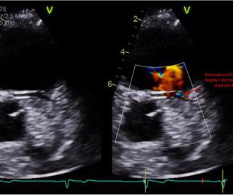
Electrocardiogram in clinic showed sinus arrhythmia with early repolarization and no ischemic changes. The echocardiogram showed normal cardiac structure and function, however, there was a concern for possible anomalous origin of the left coronary artery.
Workup including routine laboratory results, 12-lead electrocardiogram (ECG), echocardiogram, and coronary angiogram was non-specific. During the intravenous lacosamide infusion, the patient developed sudden cardiac arrest caused by ventricular arrhythmias necessitating resuscitation.
This comprehensive evaluation included the use of ultrasound echocardiograms, computed tomography (CT) scans, electrocardiograms, mutagenesis analysis, and structural analysis to gain insights into the patient's condition and the underlying mechanisms of PD.
The electrocardiogram (ECG) revealed T-wave inversions on precordial leads. Transthoracic echocardiogram (TTE) findings were consistent with Takotsubo syndrome, accompanied by mild left ventricular dysfunction. Her blood analyses demonstrated elevation of myocardial necrosis markers (peak of troponin I of 3.4
Electrocardiogram (EKG) was unremarkable. Transthoracic echocardiogram (TTE) showed an ejection fraction (EF) of 40% and a moderate-large pericardial effusion with signs of tamponade. Intra-operative transesophageal echocardiogram (TEE) post-decannulation showed a normal EF without segmental abnormalities.
Before the procedure, patients should have an electrocardiogram (ECG) and echocardiogram (ultrasound of the heart) to check the heart’s rhythm and function. Some patients need computed tomography or transoesophageal echocardiography to exclude the presence of a blood clot in the heart.
However, an echocardiogram is a different test, also conducted for heart activity. An electrocardiogram is a machine used to record the heart's electrical activity. Electrocardiogram, echocardiogram, and some other tests are done for patients with cardiac arrest. ECG and EKG refer to the same thing.
Echocardiogram is indicated (Correct) C. Start aspirin and Plavix Correct answer: (B) (B) Echocardiogram is indicated. Explanation: Shown electrocardiogram suggests left ventricular hypertrophy. Shown electrocardiogram suggests left ventricular hypertrophy. No further workup is indicated B. Start with a Free Trial.
Yes, COVID-19 symptoms can resemble a heart attack, including chest pain, shortness of breath, and changes in echocardiogram or EKG. Electrocardiograms (EKG or ECG): This diagnostic method effectively detects irregular heartbeats or arrhythmias by analyzing the electrical signals in the heart.
The attending provider wrote “Agree with electrocardiogram interpretation”. An echocardiogram showed severely reduced global systolic function with an EF of 20-25% and an LV apical thrombus. An echocardiogram showed an EF of 20-25%. The computer diagnostic algorithm diagnosed “Sinus rhythm. Normal EKG”. Normal ECG.
Electrocardiogram (ECG) and telemetry revealed junctional bradycardia with heart rate in 30s and sinus pauses (5-7 seconds). Echocardiogram was unchanged from baseline. Patient did not report any symptoms and was hemodynamically stable. He was euvolemic on physical exam. Initial laboratory analysis was unremarkable.
Laboratory tests showed markedly elevated troponin I levels (>50 ng/ml) and atrial fibrillation, along with inferior wall ST elevation on the electrocardiogram. A 2D echocardiogram revealed an ejection fraction of 43%, hypokinesia of the anterior and intraventricular septum from base to apex, and severe mitral stenosis.
Electrocardiogram (ECG) showed a prominent S wave in the left-sided leads and a prominent R wave in the right-sided chest leads, suggesting dextrocardia. His vital signs were normal, and the physical examination was unremarkable.
These tests may include: Electrocardiogram (ECG) : Records the electrical activity of your heart. Echocardiogram : Uses sound waves to create images of your heart. A heart check-up is a comprehensive evaluation of your cardiovascular health. It typically includes a physical examination, medical history review and diagnostic tests.
This is radically simplifying the patient pathway for Electrocardiogram (ECG) tests. For instance, the average waiting time for an echocardiogram at Turin’s Molinette Hospital was 31 days in 2016 and an even longer 53 days for a Holter ECG.
An initial electrocardiogram (ECG) is provided below. A rapid echocardiogram was performed, revealing an ejection fraction of 20% with thinning of the anterior-apical walls. His current medication regimen includes apixaban, carvedilol, perindopril, spironolactone, torasemide, dapagliflozin, amiodarone, and ivabradine.
Formal echocardiogram: Systolic function is at the lower limits of normal. Emergency department Code STEMI patients with initial electrocardiogram labeled ‘normal’ by computer interpretation: a 7-year retrospective review. This is a significant loss of myocardium and ejection fraction. Some function might possibly recover over weeks.
An echocardiogram on day 3 showed no wall motion abnormality (but of course, these can resolved with reperfusion, and the more time it has to resolve from "stunning", the more likely it is to be resolved). Updates on the Electrocardiogram in Acute Coronary Syndromes. It was stented. The troponin I peaked at 8.1. References : 1.
Smith , d and Muzaffer Değertekin a DIFOCCULT: DIagnostic accuracy oF electrocardiogram for acute coronary OCClUsion resuLTing in myocardial infarction. He visited an outpatient clinic for it and an echocardiogram and exercise stress test was normal. His first electrocardiogram ( ECG) is given below: --Sinus bradycardia.
Seventh , an immediate echocardiogram can make the distinction. Sixth , placement of posterior leads (take leads V4-V6 and place them at the level of the tip of the scapula, with V4 placed at the posterior axillary line ("V7"), V6 at paraspinal area ("V9"), and V5 ("V8") between them. At lease 0.5 Kligfield P, Gettes LS, Bailey JJ, et al.
Traditional tools like stethoscopes, blood pressure gauges, and electrocardiograms (ECG) are fundamental for standard diagnostic practices. This transformation extends to the use of machine learning (ML) algorithms developed by startups, which analyze medical imaging data such as ECGs, echocardiograms, and cardiac MRI scans.
Formal echocardiogram showed normal EF, no wall motion abnormalities, no pericardial effusion. Induced Brugada-type electrocardiogram, a sign for imminent malignant arrhythmias. The patient proceeded to cath where all coronaries were described as normal with no evidence of any CAD, spasm, or any other abnormality.
See this case: what do you think the echocardiogram shows in this case? New insights into the use of the 12-lead electrocardiogram for diagnosing acute myocardial infarction in the emergency department. All electrocardiograms (ECGs) and coronary angiograms were blindly analyzed by experienced cardiologists.
Formal Echocardiogram: The estimated left ventricular ejection fraction is 58 %. Emergent cardiac outcomes in patients with normal electrocardiograms in the emergency department. Because it reperfused on its own and because we intervened before it could re-occlude. Left ventricular hypertrophy concentric. Am J Emerg Med. 2021.11.023.
You will note that it is essentially an unremarkable electrocardiogram except for some PACS. Unfortunately there is no echocardiogram accessible because the patient checked himself out of the hospital in order to get back to his home state before it could be completed. In the available view, the RCA appears fully occluded.
A formal echocardiogram was completed the next day and again showed a normal ejection fraction without any focal wall motion abnormalities to suggest CAD. Induced Brugada-type electrocardiogram, a sign for imminent malignant arrhythmias. The Troponin I was cycled over time and was 0.353 followed by 0.296. Circulation, 117, 1890–1893.
Next day, a stress echo was done: The exercise stress echocardiogram is normal. The stress electrocardiogram is non-diagnostic. Normal estimated left ventricular ejection fraction improved with stress. No wall motion abnormality at rest. No wall motion abnormality with stress. The patient did not report angina with stress.
Abnormal Electrocardiogram (ECG): Defined (San Fran syncope rule) as any new changes when compared to the last ECG or presence of non-sinus rhythm. Results : Electrocardiograms (99%), telemetry (95%), cardiac enzymes (95%), and head computed tomography (CT) (63%) were the most frequently obtained tests.
His cardiac testing completed to date consist of an electrocardiogram and an echocardiogram performed Feb 16th, 2023 that were both normal. The pain resolved a few weeks later. He has had COVID twice, first in September of 2020, and his second time in January of 2023.
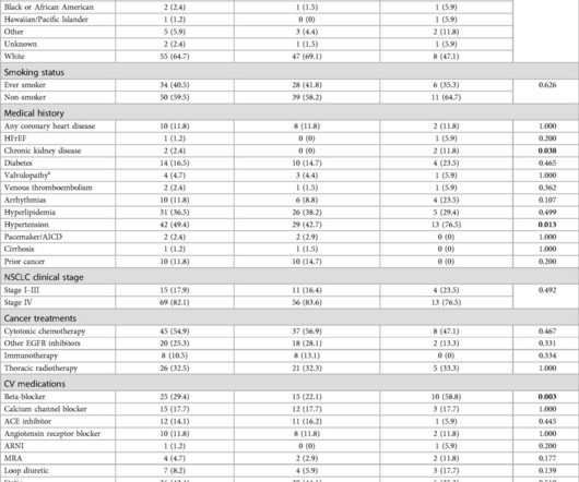
Retrospective analysis of NSCLC patients with 1 echocardiogram post-osimertinib between 2007 and 2022 was performed. Osimertinib is first-line treatment for epidermal growth factor (EGFR)-mutated non-small cell lung cancer (NSCLC) and has been associated with cardiotoxicity.
The study assessed the model's ability to analyze heart sound and single-lead electrocardiogram (ECG) data recorded via a digital stethoscope to identify individuals with actionably low EF (EF40%). Study Highlights AI's Role in Early Detection The study enrolled 2,960 adults from four U.S. healthcare networks undergoing echocardiography.
Transthoracic echocardiogram, bilateral carotid Doppler ultrasound, and electrocardiogram were normal. No previous history of hypertension or diabetes. There was no abnormality in physical examination. Cranial magnetic resonance imaging and magnetic resonance angiography showed no abnormalities.
We organize all of the trending information in your field so you don't have to. Join thousands of users and stay up to date on the latest articles your peers are reading.
You know about us, now we want to get to know you!
Let's personalize your content
Let's get even more personalized
We recognize your account from another site in our network, please click 'Send Email' below to continue with verifying your account and setting a password.


Let's personalize your content