This site uses cookies to improve your experience. To help us insure we adhere to various privacy regulations, please select your country/region of residence. If you do not select a country, we will assume you are from the United States. Select your Cookie Settings or view our Privacy Policy and Terms of Use.
Cookie Settings
Cookies and similar technologies are used on this website for proper function of the website, for tracking performance analytics and for marketing purposes. We and some of our third-party providers may use cookie data for various purposes. Please review the cookie settings below and choose your preference.
Used for the proper function of the website
Used for monitoring website traffic and interactions
Cookie Settings
Cookies and similar technologies are used on this website for proper function of the website, for tracking performance analytics and for marketing purposes. We and some of our third-party providers may use cookie data for various purposes. Please review the cookie settings below and choose your preference.
Strictly Necessary: Used for the proper function of the website
Performance/Analytics: Used for monitoring website traffic and interactions
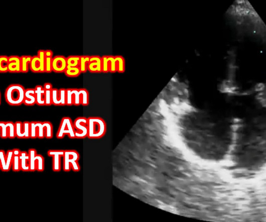
Echocardiogram in ostium primum atrial septal defect with a recap of embryological development of atrial septum. The post Echocardiogram in Ostium Primum ASD With TR appeared first on All About Cardiovascular System and Disorders.
AI can make echocardiogram analysis easier for patients to understand, according to a study published July 31 in the Journal of the American College of Cardiology: Cardiovascular Imaging.
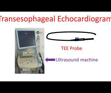
Echocardiogram is an image of the heart using ultrasound. Transesophageal echocardiogram or TEE test, is obtained by introducing a special type of transducer, also called a TEE probe, through the throat into the food pipe (esophagus) and stomach. Usual echocardiogram is obtained by placing the transducer or probe on the chest.
Fetal echocardiogram was suspicious for vascular ring with presumptive diagnosis of double aortic arch vs. circumflex right aortic arch. Post-natal echocardiogram was able to diagnose vascular ring but could not fully assess the arch or branching pattern.
A transthoracic echocardiogram was performed ( figure 1 ). Figure 1 Transthoracic echocardiogram ((A) apical four-chamber view; (B) parasternal short-axis view). The patient was referred for an exercise nuclear study and did 11 min on the Bruce protocol without angina or ischaemic ECG changes.
Transcript of the video: Now we will discuss echocardiogram in mitral valve prolapse. Billowing of the mitral leaflets is very well seen in this echocardiogram. It is a fairly common condition. Many of them may have just echo diagnosis without other relevances. Many of them may have just echo diagnosis without other relevances.
Echocardiograms using the robotic arm resulted in the same diagnosis as conventional in-person echocardiography in 98% of cases (papillary muscle level obstruction was missed in one case). tim.hodson Thu, 08/29/2024 - 11:39 Aug. 28, 2024 — New research presented at this year’s ESC Congress 2024 in London, UK (Aug. 30 – Sept.
Transcript of the video: This is a still image from a colour Doppler echocardiogram, obtained from the apical five chamber view. These are the features, you have AR jet, and MR jet, in a still image of colour Doppler echocardiogram. Left ventricle, right ventricle, left atrium and part of the aorta.
The next-day echocardiogram was made difficult by the presence in multiple projections of an acoustic shadow and abnormalities in the colour Doppler signal ( figure 1C–F ; ). The pericardial catheter was kept in place for 48 hours until complete resolution of the effusion, and then carefully removed.
Cedars-Sinai and Smidt Heart Institute investigators developed a novel foundation model that integrates computer vision interpretation of echocardiogram images with natural language processing to augment cardiologists’ interpretation of echocardiograms. Image by Getty.
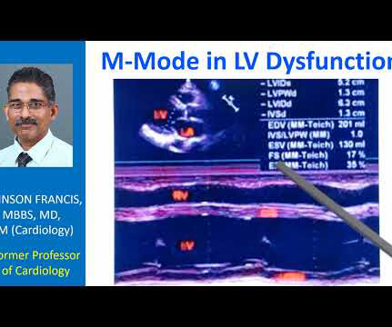
Transcript of the video: This is a still image of M-Mode Echocardiogram. M-Mode is Time-Motion Mode. The horizontal axis is time. Vertical axis is distance from the transducer. That is M-Mode, one of the older modes, currently used mainly for taking left ventricular measurements. In the inset you can see the two dimensional image.
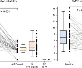
Conclusions AI can assist with the interpretation of systolic function on an echocardiogram for level 1 readers from different institutions. For the accuracy, the root-mean squared error was improved from 7.5±3.1 ±3.2 (p=0.004) p=0.004) with AI-LVEF.
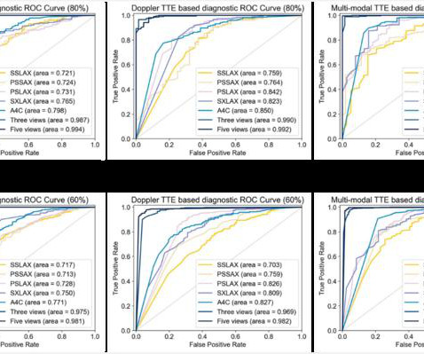
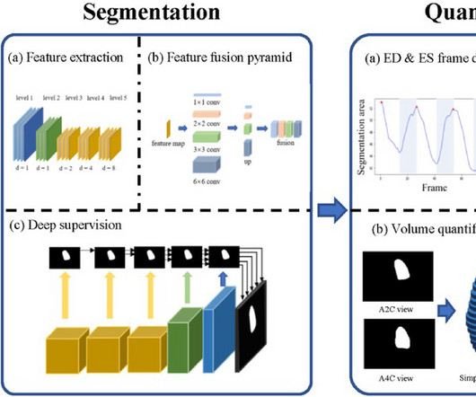
However, inexperienced sonographers often face difficulties in recognizing CHD through transthoracic echocardiogram (TTE) images. Early CHD screening and treatment can significantly improve children’s prognosis and quality of life.
Artificial intelligence experts at Cedars-Sinai and the Smidt Heart Institute created a dataset with more than 1 million echocardiograms, or cardiac ultrasound videos, and their corresponding clinical interpretations.
What do you think the echocardiogram shows? Here is the ED ECG on arrival: Less STE/STD Provider's Clinical Impression: "findings concerning for myocardial infarction, likely proximal LAD or Left main." Cath lab activated Dual antiplatelet therapy and heparin given. NTG drip started. Pain better still. First trop I returns at 1.5.
"Discover the latest guidelines from the European Society of Cardiology for managing chronic coronary syndromes, including the strong recommendation for using u
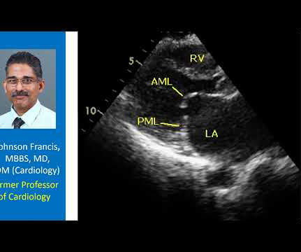
Transcript of the video: This is an apical five chamber view and this is an apical four chamber view. You can see four chambers – RV, LV, RA, LA, and the transducer location is here. And this is five chamber because, in addition you are seeing the aorta also. Right atrium has not been labelled.
A 49 year old woman with h/o COPD only presented with sudden dyspnea. She had acute pulmonary edema on exam. Prehospital Conventional algorithm interpretation: ANTERIOR INFARCT, STEMI Transformed ECG by PM Cardio: PM Cardio AI Bot interpretation: OMI with High Confidence What do you think?
Echocardiogram was performed for patients with ECMO, including at pre-ECMO, during cannulation, during ECMO support, during the ECMO wean, and a follow up within 3 months after weaning.
Echocardiogram, CT aortogram and late gadolinium imaging of the aorta have been shown in figure 1. Figure 1 (A) Two-dimensional echocardiogram short-axis basal view showing aortic valve; (B) volume-rendered CT aortogram. The patient had pregnancy-induced hypertension and hypothyroidism and was treated accordingly.
Echocardiography during pregnancy or early postpartum can assist in identifying women with preeclampsia at greater risk of future hypertension, according to a study presented at the American Society of Echocardiography's 35th Annual Scientific Sessions, held from June 14 to 16 in Portland, Oregon.
An echocardiogram (shown in a video) revealed new right ventricular dilatation, biventricular systolic dysfunction, and severe tricuspid regurgitation. Hypotension developed in a 14-year-old boy after pericardiocentesis.
Smidt Heart Institute and Cedars-Sinai, both based in Los Angeles, used the largest dataset to date to trained a machine-learning algorithm that can interpret echocardiogram images.
The importance of transesophageal echocardiogram (TEE) to exclude left atrial (LA) and left atrial appendage (LAA) thrombus prior to cardioversion for atrial fibrillation (AF) has been debated in patients who are anticoagulated prior to cardioversion. Circulation, Volume 150, Issue Suppl_1 , Page A4142210-A4142210, November 12, 2024.
Echocardiogram during that time showed stiff pulmonic valve. The patient: 4 week old female infant with past medical history of meconium aspiration at birth with APGAR scores of 2,4,6. Intubated and given nitric oxide for pulmonary hypertension. Weaned in NICU over 10 days. This ECG was obtained at follow up appointment.
The study will be launched at Cleveland Clinic and will focus on oHCM patients with the aim to validate the quality and accuracy of the monitoring echocardiograms performed by non-sonographers using UltraSights Real-Time Guidance software.
There have been limited artificial intelligence studies published assessing the potential of machine learning to detect and analyze mitral regurgitation or to detect the presence of RHD on standard portable echocardiograms.Methods and ResultsWe used 511 echocardiograms in children, focusing on color Doppler images of the mitral valve.
Developed at Children’s National Hospital and detailed in the latest edition of the Journal of the American Heart Association , the new AI system combines the power of novel ultrasound probes with portable electronic devices installed with algorithms capable of diagnosing RHD on echocardiogram.
An echocardiogram was used to confirm when the AI-enabled digital stethoscope predicted peripartum cardiomyopathy. The randomized pragmatic clinical trial enrolled 1,195 women receiving pregnancy care in Nigeria.
Research Highlights: A new software program that uses artificial intelligence to read echocardiograms may reduce the wait times for results and lead to more timely medical care. PanEcho is the first AI system to automatically assess all key areas of.
This would have been fairly easy and much more expedient to diagnose with bedside echocardiogram. The constellation of dyspnea, tachycardia, and (relatively) low voltage on ECG should prompt immediate evaluation for pericardial effusion and tamponade.
The study’s findings, published in npj Digital Medicine , suggest AI could one day be employed to analyze images from a common imaging test called an echocardiogram, which uses sound waves to capture pictures of the heart. The team trained a program to study more than 100,000 echocardiogram videos from patients with atrial fibrillation.
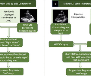
Methods A four-member expert panel reviewed 799 enrolment (in 2018) and completion (in 2020) echocardiograms from the GOAL Trial of latent RHD in Uganda to make consensus determination of normal, borderline RHD or definite RHD. Results There were 799 pairs of echocardiogram assessments included.
Transcript of the video: Closure line of aortic valve on M-Mode echocardiogram, is seen as central line, while in bicuspid aortic valve, it is an eccentric closure, nearer to one of the walls of the aorta. That is an important feature of bicuspid aortic valve on M-Mode echocardiogram. It forms almost like a box or rhomboid shape.
A test phase included 33 patients who underwent transthoracic echocardiogram with ultrasonic enhancing agent, electrocardiogram, and vectorcardiogram (aneurysm group - n = 22, and akinesia group - n = 11). The medical records of 2,670 individuals were analyzed in this cross-sectional study.
However, there is high interobserver variability with visual estimation methods, which can be compounded by inadequate quality of capture and lack of specialised training in both obtaining and interpreting echocardiograms in these settings.
Our report describes two cases of SVS treated with endocardial ablation to improve LVOTO.Case reportCase 1: A 74-year-old female patient with angina and syncope was admitted to the hospital and diagnosed with SVS by transthoracic echocardiogram. After RFA was performed, the patient's symptoms significantly improved.
A transthoracic echocardiogram demonstrated a large left atrial mass extending into the right upper pulmonary veins. A 73-year-old woman presented to the emergency department with a syncopal episode and a history of dizzy spells.
This study investigates the correlation of left atrial enlargement and left ventricular hypertrophy (LVH) found on ECG with transthoracic echocardiogram and cardiac magnetic resonance imaging (CMR) findings.MethodsThis is a singlecenter retrospective study of 1000 patients. CMR data were included if within 6 months of the ECG.
Cardiac implantable electronic device(CIED) extraction is recommended in patients with related infective endocarditis (IE). Patients with lead-associated vegetation larger than 10 mm face increased odds of embolism and mortality. Percutaneous mechanical aspiration devices reduce the risk of embolization during lead extraction.
We organize all of the trending information in your field so you don't have to. Join thousands of users and stay up to date on the latest articles your peers are reading.
You know about us, now we want to get to know you!
Let's personalize your content
Let's get even more personalized
We recognize your account from another site in our network, please click 'Send Email' below to continue with verifying your account and setting a password.


Let's personalize your content