This site uses cookies to improve your experience. To help us insure we adhere to various privacy regulations, please select your country/region of residence. If you do not select a country, we will assume you are from the United States. Select your Cookie Settings or view our Privacy Policy and Terms of Use.
Cookie Settings
Cookies and similar technologies are used on this website for proper function of the website, for tracking performance analytics and for marketing purposes. We and some of our third-party providers may use cookie data for various purposes. Please review the cookie settings below and choose your preference.
Used for the proper function of the website
Used for monitoring website traffic and interactions
Cookie Settings
Cookies and similar technologies are used on this website for proper function of the website, for tracking performance analytics and for marketing purposes. We and some of our third-party providers may use cookie data for various purposes. Please review the cookie settings below and choose your preference.
Strictly Necessary: Used for the proper function of the website
Performance/Analytics: Used for monitoring website traffic and interactions
Here is her ECG: Regular Wide Complex Tachycardia. Could it be atrial tachycardia with RBBB and LPFB aberrancy? Here it is: There is sinus with normal conduction, very different from her tachycardia. Severely decreased LV function. What do you think? What do you want to do? She was not on any medication that could cause this.
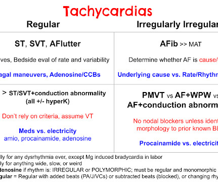
ECG#1 There is a regular tachycardia with a ventricular rate of about 180 bpm. Smith comment : When there is a regular wide complex tachycardia, first assess whether it is sinus or not. Is it sinus or is it a supraventricular dysrhythmia? Put shortly is SVT with "Shark Fin STE" and not ventricular tachycardia.
The ECGs show a wide complex, irregularly irregular tachycardia. At that time, he presented via EMS and had received magnesium and lidocaine prehospital for concerns of ventricular tachycardia. On arrival to the ED, he was noted to be in a wide complex tachycardia with a rate in the 240s.
Because she has cardiomyopathy and ventricular dysrhythmias, the pacer included an Implanted Cardioverter-Defibrillator (ICD) Echo 6 days later after CRT: Normal estimated left ventricular ejection fraction. Even with tachycardia and a paced QRS duration of ~0.16 No wall motion abnormality. (J J Am Coll Cardiol.
A young male with unknown past medical history presents with AMS and tachycardia. There is sinus tachycardia, a prolonged QRS (computer read it as 114 ms, previous ECG with 102 ms). No patient with a QRS of less than 160 ms had ventricular dysrhythmias. The preshospital ECG and strips are not available.
ECG at presentation was suggestive of ventricular tachycardia (VT) ( figure 1 A ). Resuscitation with urgent cardioversion in view of haemodynamic instability with wide complex tachycardia was done. On examination, the pulse rate was around 190 beats/min with a systolic blood pressure of 80 mm Hg.
The ECG shows sinus tachycardia with RBBB and LAFB, without clear additional superimposed signs of ischemia. See these publications for more information Overall, management for cardiac contusion is mostly supportive unless surgical complications develop, involving appropriate treatment of dysrhythmias and hemodynamic instability.
Competitive atrial pacing (CAP) is a pacemaker-mediated dysrhythmia that leads to functional atrial undersensing and loss of capture. This condition may mimic pacemaker-mediated tachycardia (PMT) symptoms and has the potential to induce atrial fibrillation (AF).
They had already cardioverted at 120 J, then 200 J, which resulted in the following: Ventricular Tachycardia They then cardioverted at 200 J which r esulted in the same narrow complex rhythm shown above, at 185 beats per minute. This would treat both SVT or sinus tachycardia. I suggested esmolol if the heart rate did not improve.
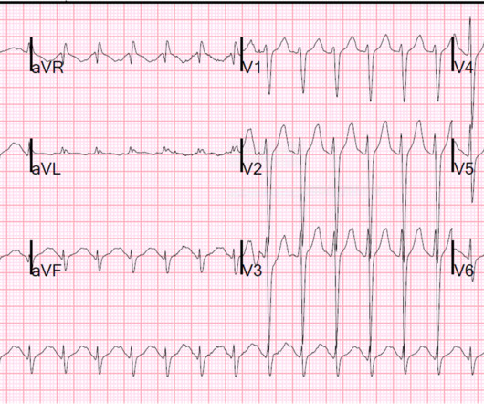
Here is her ED ECG: Here is the ED physician's interpretation: IMPRESSION UNCERTAIN REGULAR RHYTHM, wide complex tachycardia, likely p-waves. LEFT BUNDLE BRANCH BLOCK [120+ ms QRS DURATION, 80+ ms Q/S IN V1/V2, 85+ ms R IN I/aVL/V5/V6] Comparison Summary: LBBB and tachycardia are new. This is clearly ventricular tachycardia.
Multifocal Atrial Tachycardia 2. Atrial dysrhythmias, and atrial fi brillation in particular, are frequently misdiagnosed by computer algorithms and then by the physician who overreads them. The rhythm is indeed irregularly irregular, so atrial fibrillation must be considered. Sinus with multifocal PACs 3. Sinus with multifocal PVCs 4.
We see a regular tachycardia with a narrow QRS complex and no evidence of OMI or subendocardial ischemia. The differential of a regular narrow QRS tachycardia is sinus tachycardia, SVT, and atrial flutter with regular conduction. Now the patient is in sinus tachycardia. Her initial EKG is below. Same as initial ECG.
Interpretation: There is sinus tachycardia, with right bundle branch block (RBBB). Blunt cardiac injury my result in : 1) Acute myocardial rupture with tamponade 2) Valve rupture (tricuspid, aortic, mitral) 3) Coronary thrombosis or dissection (and thus Acute MI) from direct coronary blunt injury 4) Dysrhythmias of all kinds.
Tachycardia and ST Elevation. Likewise, in some cases of ischemia concealed by flutter waves, the ischemia can be seen despite the flutter waves, whereas in other cases the dysrhythmia must be terminated before the ischemia can be clearly distinguished. Tachycardia to this degree can cause ST segment changes in several ways.
Our electrophysiologist, Rehan Karim, states he has ablated AVNR"T" ("T" because it is not tachycardia) in a 90 year old, and that he has seen rate-related BBB at very slow rates. The second explanation (AIVR), whether as a reperfusion dysrhythmia or not, seems most likely.
Here is his 12-lead: There is a wide complex tachycardia with a rate of 257, with RBBB and LPFB (right axis deviation) morphology. Read about Fascicular VT here: Idiopathic Ventricular Tachycardias for the EM Physician Case Continued He was completely stable, so adenosine was administered. See Learning point 1 below. Arch Intern Med.
Here is the ECG: Sinus tachycardia. So the real QT is shorter, but the computer does not mention the U-wave, and the U-wave is as important as the T-wave in predicting cardiac dysrhythmias. This patient presented with severe DKA. What do you think? The computer and physician reader wrote: "ST depression, consider subendocardial injury."
Is it ventricular tachycardia (VT) due to hyperK or is it a supraventricular rhythm with hyperK? Here are other posts on hyperK, large calcium doses for hyperK, and ventricular tachycardia in hyperK Weakness, prolonged PR interval, wide complex, ventricular tachycardia Very Wide and Very Fast, What is it? How would you treat?
Opinions vary widely on the K level at which a patient must be admitted on a monitor because of the risk of ventricular dysrhythmias. My rationale is that if the K is affecting the ECG, then it is affecting the electrical milieu and can result in serious dysrhythmias. Until some real data is available, my opinion is this: 1.
I find AV dissociation in VT to be very difficult to differentiate from artifact, as there are always random blips on tachycardia tracings. If you don't know what the dysrhythmia is, then try procainamide. Read this post: Idiopathic Ventricular Tachycardias for the EM Physician 2. Ken notes AV dissociation. What to do now?
Smith comment: In a large randomized trial of dopamine vs. norepinephrine (11) for shock which was published after the above-mentioned recommendations, dopamine had more adverse events (especially severe dysrhythmias, and especially atrial fibrillation). Hypotension may of course be a result of a brady- or tachydysrhythmia.
Here was his ED ECG: There is sinus tachycardia (rate about 114) with nonspecific ST-T abnormalities. An ECG was recorded: This shows a regular narrow complex tachycardia at a rate of about 160. See my quick review of atrial tachycardia below) The tachycardia spontaneously resolved. BP:143/99, Pulse 109, Temp 37.2 °C
See here for management of Polymorphic Ventricular Tachycardia , which includes Torsades. Could the dysrhythmias have been prevented? Severe hypokalemia in the setting of STEMI or dysrhythmias is life-threatening and needs very rapid treatment. Learning Points: 1. 5-10 mEq over 5-10 minutes is appropriate for a K of 1.8
Then there is loss of pulses with continued narrow complex on the monitor ("PEA arrest") Learning Points: Sudden witnessed Cardiac Arrest due to ACS is almost always due to dysrhythmia. Tachycardia is of course, quite common in patients following cardiac arrest.
Otherwise vitals after intubation were only notable for tachycardia. An initial EKG was obtained: Computer read: sinus tachycardia, early acute anterior infarct. She was ventilated by bag-valve-mask by EMS on arrival and was quickly intubated with etomidate and succinylcholine. A rectal temperature was obtained which read 107.9
If the patient has Abnormal Vital Signs (fever, hypotension, tachycardia, or tachypnea, or hypoxemia), then these are the primary issue to address, as there is ongoing pathology which must be identified. Most physicians will automatically be worried about these symptoms. The tracings were considered abnormal in the following cases: 1.
Smith comments : Wide complex tachycardia. The differential diagnosis of WCT is: 1) Sinus tachycardia with "aberrancy" (in this case RBBB and LAFB), but there are no P-waves and the QRS morphology is not typical of simple RBBB/LAFB. Also, if the rate is constant, not wavering up and down, it is highly unlikely to be sinus tachycardia.
We organize all of the trending information in your field so you don't have to. Join thousands of users and stay up to date on the latest articles your peers are reading.
You know about us, now we want to get to know you!
Let's personalize your content
Let's get even more personalized
We recognize your account from another site in our network, please click 'Send Email' below to continue with verifying your account and setting a password.


Let's personalize your content