This site uses cookies to improve your experience. To help us insure we adhere to various privacy regulations, please select your country/region of residence. If you do not select a country, we will assume you are from the United States. Select your Cookie Settings or view our Privacy Policy and Terms of Use.
Cookie Settings
Cookies and similar technologies are used on this website for proper function of the website, for tracking performance analytics and for marketing purposes. We and some of our third-party providers may use cookie data for various purposes. Please review the cookie settings below and choose your preference.
Used for the proper function of the website
Used for monitoring website traffic and interactions
Cookie Settings
Cookies and similar technologies are used on this website for proper function of the website, for tracking performance analytics and for marketing purposes. We and some of our third-party providers may use cookie data for various purposes. Please review the cookie settings below and choose your preference.
Strictly Necessary: Used for the proper function of the website
Performance/Analytics: Used for monitoring website traffic and interactions
Clinician and EKG machine read of acute pericarditis. While it is true that inferior MI has ST depression in aVL 99% of the time (Bischof and Smith), and that inferolateral ST elevation is the most common distribution for pericarditis, the ST elevation in V3 has "terminal QRS distortion (TQRSD)," (diagnostic of LAD occlusion).
After admission he undergoes another ECG, though it is unclear from documentation whether there was a change in his chest pain. Remember, pericarditis is the thing you say and write down when youre actively trying to miss an OMI. Remember, pericarditis is the thing you say and write down when youre actively trying to miss an OMI.
There is a reasonable chance of pericarditis in this case, or this could be a baseline." Sadly, I did not receive enough information to adjudicate whether this patient has pericarditis or not. I sent this to Dr. Smith and this was his response: "Likely pericarditis, but that is perilous. I immediately responded: "cool fake!
Tuberculous pericardial effusion can be documented by aspirating the fluid and culturing the fluid for the presence of the bacteria causing tuberculosis (Mycobacterium tuberculosis). There are other tests also for tuberculous pericarditis, but they not as sure as growing the bacterium in culture.
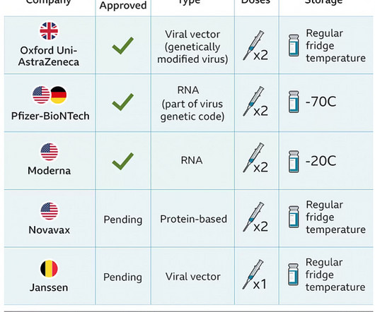
The case reports Case 1 involves a 26 year old man who developed pericarditis after the Pfizer vaccine. Pericarditis, an inflammation of the sac the heart lives in, developed about 7 days after the Pfizer vaccine. Abnormal cardiac biomarkers suggested damage to the heart muscle confirming myocarditis.
Haven't you been taught that this favors pericarditis? Weren't you taught that concave morphology favors pericarditis? Expert ECG interpretation can often distinguish normal variant STE from OMI from pericarditis. Smith = “You diagnose acute pericarditis at your peril”. We will study this soon with our database.
Below is the first ECG, signed off by the over-reading cardiologist agreeing with the computer interpretation: ST elevation, consider early repolarization, pericarditis, or injury. Theres ST elevation in V3-4 which meets STEMI criteria, which could be present in either early repolarization, pericarditis or injury. What do you think?
Echo studies in patients with documented cardiac tamponade confirm that electrical alternans is synchronous with and a direct result of the pendulous movement of the heart within the enlarged, fluid-filled pericardial sac of a patient with large pericardial effusion ( the "swinging heart" phenomenon ).
This is a bad ST vector orientation, because it causes widespread STE and one of the most important mistakes that needs to be avoided here is thinking of the diagnosis of pericarditis. Such an out-of-proportion STE is virtually never seen in pericarditis. Look at the STE in lead II, aVF. Smith's Blog show this same phenomenon ).
Pericarditis? A straight ST segment virtually never happens in inferior ST elevation that is NOT due to OMI (normal variant, pericarditis) 4. In this patient with documented coronary disease — these q waves could reflect prior lateral infarction ( especially in view of the Q in lead aVL ). Time zero What do you think?
The second most common cause of medical cardiac tamponade is acute idiopathic pericarditis. Less common etiologies include uremia, bacterial or tubercular pericarditis, chronic idiopathic pericarditis, hemorrhage, and other causes such as autoimmune diseases, radiation, myxedema, etc.
You can easily imagine this patient getting one of several diagnoses -- vasospasm, MINOCA , pericarditis, or maybe even no diagnosis at all beyond "non-obstructive coronary artery disease." The operator documented thoughtful consideration of risks and benefits of stent placement. At the time of IVUS, there was no thrombus.
This prompted a repeat ECG (we do not have documentation from that time to tell us whether he had persistent, recurrent, or absent pain): Progression of anterior OMI to full Q-wave MI with large pathologic Q-waves in V2-V4 with persistent STE which now meets STEMI criteria (after full thickness infarction/stunning). ng/mL (very elevated).
Acute procedure success (defined as confirmed entrance block at the end of procedure) and freedom from documented atrial arrhythmia recurrence at 12 months were also assessed. About the VARIPULSE Platform The VARIPULSE Platform is Biosense Webster's Irreversible Electroporation ablation system.
Triage documented a complaint of left shoulder pain. Recall from this post referencing this study that "reciprocal STD in aVL is highly sensitive for inferior OMI (far better than STEMI criteria) and excludes pericarditis, but is not specific for OMI." The patient presented to triage at around 10 PM. link] Bischof, J. Worrall, C.,
Dyspnea, Chest pain, Tachypneic, Ill appearing: Bedside Cardiac Echo gives the Diagnosis 31 Year Old Male with RUQ Pain and a History of Pericarditis. Submitted by a Med Student, with Great Commentary on Bias! Chest pain, SOB, Precordial T-wave inversions, and positive troponin. What is the Diagnosis?
They include myocardial ischemia, acute pericarditis, pulmonary embolism, external compression due to mass over the right ventricular outflow tract region, and metabolic disorders like hyper or hypokalemia and hypercalcemia. These are the conditions which have to be considered or excluded as they can sometimes manifest Brugada pattern on ECG.
The emergency medicine physician documented, "His initial EKG is riddled with artifact and difficult to interpret but does not look like a STEMI." Several hours passed with no documentation as to the reason for delay. In fact, even the GE algorithm got this one (partially) right.
We organize all of the trending information in your field so you don't have to. Join thousands of users and stay up to date on the latest articles your peers are reading.
You know about us, now we want to get to know you!
Let's personalize your content
Let's get even more personalized
We recognize your account from another site in our network, please click 'Send Email' below to continue with verifying your account and setting a password.


Let's personalize your content