This site uses cookies to improve your experience. To help us insure we adhere to various privacy regulations, please select your country/region of residence. If you do not select a country, we will assume you are from the United States. Select your Cookie Settings or view our Privacy Policy and Terms of Use.
Cookie Settings
Cookies and similar technologies are used on this website for proper function of the website, for tracking performance analytics and for marketing purposes. We and some of our third-party providers may use cookie data for various purposes. Please review the cookie settings below and choose your preference.
Used for the proper function of the website
Used for monitoring website traffic and interactions
Cookie Settings
Cookies and similar technologies are used on this website for proper function of the website, for tracking performance analytics and for marketing purposes. We and some of our third-party providers may use cookie data for various purposes. Please review the cookie settings below and choose your preference.
Strictly Necessary: Used for the proper function of the website
Performance/Analytics: Used for monitoring website traffic and interactions
Patients had routine 12-lead electrocardiograms (ECGs) regardless of presenting complaints. Data regarding AF screening in conflict countries emergency departments (ED) is lacking.MethodsWe included consecutive patients >40 years old who reported to the ED of a Syrian tertiary centre between July 2024 and September 2024.
Diabetic Cardiomyopathy (DCM) is a diabetes mellitus-induced pathophysiological condition that can lead to heart failure. Diabetic rats were established by injection of streptozotocin (STZ, 6085 mg/kg). Diabetic Cardiomyopathy (DCM) is a diabetes mellitus-induced pathophysiological condition that can lead to heart failure.
Recent guidelines propose N-terminal pro-B-type natriuretic peptide (NT-proBNP) for recognition of asymptomatic left ventricular (LV) dysfunction (Stage B Heart Failure, SBHF) in type 2 diabetes mellitus (T2DM.
Background:GLP-1 receptor agonists (GLP1RA) agonists have been shown to reduce cardiovascular events in patients with type 2 diabetes (T2D) and atherosclerotic cardiovascular disease (ASCVD). However, it is unclear whether early subclinical therapeutic changes can be detected from routine 12-lead electrocardiograms (ECGs).Methods:We
Electrocardiogram (ECG) and telemetry revealed junctional bradycardia with heart rate in 30s and sinus pauses (5-7 seconds). Patient did not report any symptoms and was hemodynamically stable. He was euvolemic on physical exam.
His medical history includes hypertension, a decade-long battle with diabetes, ischemic heart disease, a coronary bypass graft surgery ten years ago, a diagnosis of congestive heart failure for the last five years, and a prior ICD implantation five years ago. An initial electrocardiogram (ECG) is provided below. What do you think?
This 57 yo diabetic male presented with generalized fatigue, myalgias, and arthralgias , mild subjective fever and chills, and nausea. This 42 yo diabetic male presented with cough and foot pain. Clinical value of 12-lead electrocardiogram after successful reperfusion therapy for acute myocardial infarction.
Using digital cardiac care tools resulted in an increase considering their positive role in monitoring health through: Smoking cessation Weight loss programs Optimised blood pressure control Glycemic control in diabetes Lipid and cholesterol levels Recommended Read: Medical Imaging Emerging Trends in 2021 2.
Traditional tools like stethoscopes, blood pressure gauges, and electrocardiograms (ECG) are fundamental for standard diagnostic practices. Much of heart disease is linked to conditions like diabetes, obesity, or chronic kidney disease, and specific mutations in nucleic acids help identify them.
This was a male in his 50's with a history of hypertension and possible diabetes mellitus who presented to the emergency department with a history of squeezing chest pain, lasting 5 minutes at a time, with several episodes over the past couple of months. Also see this incredible case of the use of 12-lead ST Segment monitoring.
Case submitted and written by Mazen El-Baba MD, with edits from Jesse McLaren and edits/comments by Smith and Grauer A 90-year old with a past medical history of atrial fibrillation, type-2 diabetes, hypertension, dyslipidemia, presented with acute onset chest/epigastric pain, nausea, and vomiting. BP was 110 and oxygen saturation was normal.
This results in severe chest pain or discomfort, with the subsequent release of cardiac biomarkers, and alterations in the electrocardiogram. Hypertension and diabetes were the two most common risk factors identified. It can cause diminished heart function and mortality if not treated properly with suitable measures.
Electrocardiogram (ECG/EKG) An ECG records the electrical activity of the heart and can help detect abnormalities in the heart’s rhythm that might contribute to enlargement. Monitor cholesterol levels and manage conditions like diabetes that can strain the heart. Exercise regularly to keep the heart strong and healthy.
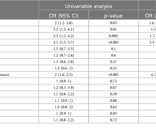
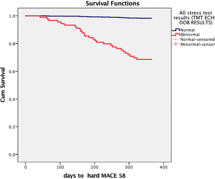
Background Patients with low HEART (History, Electrocardiogram, Age, Risk factors, and Troponin level) risk scores who are discharged from the emergency department (ED) may present clinical challenges and diagnostic dilemmas. The use of downstream non-invasive stress imaging (NISI) tests in this population remains uncertain.
Moreover, electrocardiograms, which record the electrical activity of the heart, and wearable devices can provide artificial intelligence (AI) the data it needs to spot possible cases of valvular heart disease via fluctuations in heart rate, blood pressure, blood oxygenation and other factors.
These tests may include: Electrocardiogram (ECG) : Records the electrical activity of your heart. A heart check-up is a comprehensive evaluation of your cardiovascular health. It typically includes a physical examination, medical history review and diagnostic tests. Echocardiogram : Uses sound waves to create images of your heart.
While factors like high blood pressure, high cholesterol, obesity, and smoking affect both men and women, certain conditions like diabetes, metabolic syndrome, and mental stress tend to pose a higher risk for women. Hormonal changes, particularly during menopause, also play a significant role, altering women’s cardiovascular health.
Background:In patients with type 2 diabetes (T2D), hyperglycemia and glycemic variability lead to prolongation and greater heterogeneity of ventricular repolarization, manifested on the electrocardiogram through an increase in QT, QTc, TpeakTend (TpTe) intervals and the TpTe/QT ratio, increasing the risk of potentially malignant arrhythmias.
An artificial intelligence-based electrocardiogram (ECG) AF algorithm (AI-AF) can effectively identify silent AF. Introduction:Patients with atherosclerotic carotid artery disease are at high risk of mortality in the long-term follow-up after carotid endarterectomy (CEA), partly due to dysrhythmia.
LR was based on normal examination, stable hemodynamics, normal electrocardiograms (ECG), and negative cardiac troponin I, without pre-discharge functional or anatomic cardiac testing or risk scores. Length of stay (LOS) in the CPU to discharge was 10.4
Edits by Meyers and Smith A man in his 70s with PMH of hypertension, hyperlipidemia, type 2 diabetes, CVA, dual-chamber Medtronic pacemaker, presented to the ED for evaluation of acute chest pain. Sent by Pete McKenna M.D. Triage ECG: What do you think? This is diagnostic of proximal LAD occlusion. This is a huge anterolateral OMI.
Written by Jesse McLaren An 80 year old patient with diabetes/hypertension/ cirrhosis had a recent increase in candesartan for their hypertension, and was also on spirolactone and nadolol. Severe hyperkalemia: can the electrocardiogram risk stratify for short-term adverse events. Labs showed a non-hemolyzed potassium of 6.2,
No previous history of hypertension or diabetes. Transthoracic echocardiogram, bilateral carotid Doppler ultrasound, and electrocardiogram were normal. There was no abnormality in physical examination. Cranial magnetic resonance imaging and magnetic resonance angiography showed no abnormalities.
We organize all of the trending information in your field so you don't have to. Join thousands of users and stay up to date on the latest articles your peers are reading.
You know about us, now we want to get to know you!
Let's personalize your content
Let's get even more personalized
We recognize your account from another site in our network, please click 'Send Email' below to continue with verifying your account and setting a password.


Let's personalize your content