This site uses cookies to improve your experience. To help us insure we adhere to various privacy regulations, please select your country/region of residence. If you do not select a country, we will assume you are from the United States. Select your Cookie Settings or view our Privacy Policy and Terms of Use.
Cookie Settings
Cookies and similar technologies are used on this website for proper function of the website, for tracking performance analytics and for marketing purposes. We and some of our third-party providers may use cookie data for various purposes. Please review the cookie settings below and choose your preference.
Used for the proper function of the website
Used for monitoring website traffic and interactions
Cookie Settings
Cookies and similar technologies are used on this website for proper function of the website, for tracking performance analytics and for marketing purposes. We and some of our third-party providers may use cookie data for various purposes. Please review the cookie settings below and choose your preference.
Strictly Necessary: Used for the proper function of the website
Performance/Analytics: Used for monitoring website traffic and interactions
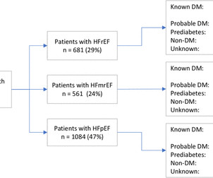
Aims The aim of this study was to investigate a real-world heart failure (HF) cohort regarding (1) prevalence of known diabetes mellitus (DM), undiagnosed DM and pre-diabetes, (2) if hf treatment differs depending on glycaemic status and (3) if treatment of DM differs depending on HF phenotype.
Pulse was 115, BP 140/65, and afebrile He was found to have cellulitis and to be in diabetic ketoacidosis, with bicarb of 14, pH of 2.27, glucose of 381, anion gap of 18, and lactate of 2.2 See this post: What do you think the echocardiogram shows in this case? He was treated for infection and DKA and admission to hospital was planned.
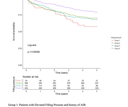
However, the impact of elevated FP as detected by pretranscatheter aortic valve replacement (TAVR) echocardiogram on long-term outcomes after TAVR remains unclear. The presence of elevated FP was determined in accordance with the latest guidelines using the last available comprehensive echocardiogram prior to TAVR.
Echocardiogram was unchanged from baseline. Patient did not report any symptoms and was hemodynamically stable. His home medications included metoprolol succinate 25mg daily which was held given bradycardia. Urine drug screen was positive for cannabis.
A 56 year old male with a history of diabetes, dyslipidemia, hypertension, and coronary artery disease presented to the emergency department with sudden onset weakness, fatigue, lethargy, and confusion. This is another case sent by the undergraduate (who is applying to med school) who works as an EKG tech. No ECG was ordered on Day #1.
Residual shunt post PFO closure was assessed using transthoracic echocardiogram (TTE) with saline contrast. These findings remained robust after adjusting for other VCID risk factors, such as age, diabetes, hyperlipidemia and hypertension (Table 1).Conclusion:Our All the patients were cognitively normal at the time of PFO diagnosis.
Advanced cardiac workup (ACW), including transesophageal echocardiogram (TEE) and implantable loop recorder (ILR) are widely considered a crucial element in the ESUS work-up. Baseline characteristics such as diabetes, hypertension, smoking, ipsilateral ICAD, etc. The etiology of AChA infarcts remains poorly understood. IQR of 13.5)
Identified echocardiograms were reviewed to confirm IVMS aneurysms and exclude sinus of Valsalva aneurysms. One patient had diabetes, 8 had hyperlipidemia, and 9 had hypertension. Patients with concurrent structural heart anomalies were excluded. Of these, 9 (64%) were female with a mean age of 59.6, The mean BMI was 27.9
His medical history includes hypertension, a decade-long battle with diabetes, ischemic heart disease, a coronary bypass graft surgery ten years ago, a diagnosis of congestive heart failure for the last five years, and a prior ICD implantation five years ago. The initial troponin T level was measured at 30 ng/L.
Dysrhythmias are more likely in patients who are older and sicker with a larger burden of comorbidities such as diabetes, high blood pressure, sleep apnoea and vascular disease. There are two other important points to note. Most dysrhythmias once identified are easily treatable.
link] A 62 year old man with a history of hypertension, type 2 diabetes mellitus, and carotid artery stenosis called 911 at 9:30 in the morning with complaint of chest pain. His echocardiogram showed normal wall motion. This is written by Willy Frick, an amazing cardiology fellow in St. Before and after of the LAD shown below.
This transformation extends to the use of machine learning (ML) algorithms developed by startups, which analyze medical imaging data such as ECGs, echocardiograms, and cardiac MRI scans. Much of heart disease is linked to conditions like diabetes, obesity, or chronic kidney disease, and specific mutations in nucleic acids help identify them.
female with HTN, HLD, diabetes, ESRD on dialysis is brought in by EMS with sudden onset, left -sided chest pain for the past four hours. Given her risk factors (HTN, HLD, ESRD from diabetes) I decided to obtain a broad cardiac workup for the patient: serial ECGs, labs, serial troponins, CXR and bedside cardiac ultrasound.
This was a male in his 50's with a history of hypertension and possible diabetes mellitus who presented to the emergency department with a history of squeezing chest pain, lasting 5 minutes at a time, with several episodes over the past couple of months. Also see this incredible case of the use of 12-lead ST Segment monitoring.
A 40-something woman with diabetes and peripheral vascular disease who frequently needs the ED for chronic pain called 911 for sudden severe chest pain. Echocardiogram: The estimated left ventricular ejection fraction is 34% Regional wall motion abnormality-lateral, akinetic. The patient was very agitated and could not hold still.
Case submitted and written by Mazen El-Baba MD, with edits from Jesse McLaren and edits/comments by Smith and Grauer A 90-year old with a past medical history of atrial fibrillation, type-2 diabetes, hypertension, dyslipidemia, presented with acute onset chest/epigastric pain, nausea, and vomiting. BP was 110 and oxygen saturation was normal.
Echocardiogram An echocardiogram uses sound waves to produce a detailed image of the heart, allowing doctors to see the size of the heart chambers and how well the heart is pumping blood. Monitor cholesterol levels and manage conditions like diabetes that can strain the heart. The following diagnostic tools are commonly used: 1.
Maintain a Healthy Weight: Obesity amplifies the effects of genetic predispositions by contributing to high cholesterol, hypertension, and diabetes. Heart imaging, such as echocardiograms or CT scans. Medication and Medical Interventions Sometimes, lifestyle changes alone may not be enough to counteract genetic risks.
The presence of type 2 diabetes not only signifies a chronic metabolic disorder, but also serves as a catalyst for various cardiovascular and cerebrovascular ailments such as coronary heart disease and stroke.
Our objective was to evaluate the prevalence of DD in people living with human immunodeficiency virus without known history of diabetes or hypertension in Western Kenya. Study participants underwent a comprehensive two-dimensional echocardiogram and laboratory testing. Despite low prevalence of DD, PLWH had 5.76
Echocardiogram : Uses sound waves to create images of your heart. It typically includes a physical examination, medical history review and diagnostic tests. These tests may include: Electrocardiogram (ECG) : Records the electrical activity of your heart. Stress test : Assesses your heart’s function under stress.
ARIES-HM3 Subgroup Analysis: Nir Uriel, MD (USA) presented findings from a subgroup analysis of ARIES-HM3, showing that avoiding aspirin in newly implanted HeartMate 3 LVAD patients with atrial fibrillation, diabetes, and obesity significantly reduces non-surgical bleeding events at one year while maintaining safety.
This was sent to me by a French colleague, Olivier Peyronie "Yesterday we received a 62 yo man with diabetes, hypertension and smoker. The echocardiogram shows a preserved left ventricular ejection fraction (LVEF) of 55% with marked basal and mid inferolateral and basal anterolateral hypokinesia. Time zero: What do you think?
A patient in their 40s with type 1 diabetes mellitus and hyperlipidemia presented to the emergency department with 5 days of “flu-like” illness. While awaiting transfer to the cath lab, STAT echocardiogram was performed and showed LVEF 30-35%, as well as anterior, inferior, and apical hypokinesis, and apical thrombus.
It is also very important to mention a history of high blood pressure, diabetes, elevated cholesterol, family history of premature heart disease, stroke or even sudden death. Another way of imaging the heart is via a transesophageal echocardiogram.
All underwent MRI to measure coronary vessel wall thickness and an echocardiogram to assess left ventricular function. senior clinical investigator, and director of the Biomedical and Metabolic Imaging Branch, National Institute of Diabetes and Digestive and Kidney Diseases ( NIDDK ), in Bethesda, Maryland. Gharib, M.D.
Written by Willy Frick A man in his mid 30s with type 1 diabetes presented with two days of midsternal and epigastric pain, described as both "sharp" and squeezing." This is a very bold statement in a type 1 diabetic with very concerning sounding chest pain. Echocardiogram showed LVEF 33% with akinesis of the lateral wall.
No previous history of hypertension or diabetes. Transthoracic echocardiogram, bilateral carotid Doppler ultrasound, and electrocardiogram were normal. There was no abnormality in physical examination. Cranial magnetic resonance imaging and magnetic resonance angiography showed no abnormalities.
We organize all of the trending information in your field so you don't have to. Join thousands of users and stay up to date on the latest articles your peers are reading.
You know about us, now we want to get to know you!
Let's personalize your content
Let's get even more personalized
We recognize your account from another site in our network, please click 'Send Email' below to continue with verifying your account and setting a password.


Let's personalize your content