This site uses cookies to improve your experience. To help us insure we adhere to various privacy regulations, please select your country/region of residence. If you do not select a country, we will assume you are from the United States. Select your Cookie Settings or view our Privacy Policy and Terms of Use.
Cookie Settings
Cookies and similar technologies are used on this website for proper function of the website, for tracking performance analytics and for marketing purposes. We and some of our third-party providers may use cookie data for various purposes. Please review the cookie settings below and choose your preference.
Used for the proper function of the website
Used for monitoring website traffic and interactions
Cookie Settings
Cookies and similar technologies are used on this website for proper function of the website, for tracking performance analytics and for marketing purposes. We and some of our third-party providers may use cookie data for various purposes. Please review the cookie settings below and choose your preference.
Strictly Necessary: Used for the proper function of the website
Performance/Analytics: Used for monitoring website traffic and interactions
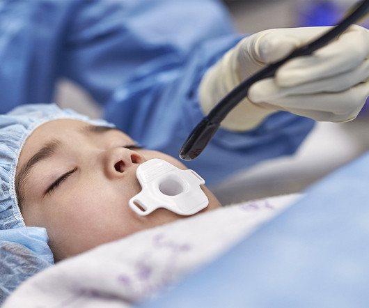
Cardiovascular ultrasound has played a key role in the evolution of early diagnosis of structural heart disease, led by a technology pioneered by Philips: the ‘transesophageal echocardiography’ (TEE) ultrasound transducer. TEE helps cardiologists by providing highly detailed images of the heart and its internal structures.
Food and Drug Administration (FDA) has granted 510(k) clearance for its first-of-a-kind, AI-powered AISAP CARDIO point-of-careultrasound (POCUS) software platform. Prospective data have shown that use of CARDIO has generated improvement in clinician performance, with care pathway changes in one third of scanned patients.
"At GE HealthCare, we understand the critical challenges healthcare providers face, from staffing shortages to complex workflows. Our AI-enabled portfolio, including our Command Center Software Platform, Edison True PACS , and Venue Family ultrasound systems with Caption Guidance , is designed to directly address these issues.
Arrival at time 0 ECG 7 min Roomed in hallway at 17 min Moved to room with monitor at 37 min The patient was seen briefly by the physician, who then went to get an ultrasound machine. Then the patient would have been taken to the criticalcare area with a defibrillator at his side while waiting for the cath lab to be ready.
He was rushed by residents into our criticalcare room with a diagnosis of STEMI, and they handed me this ECG: There is sinus tachycardia with ST elevation in II, III, and aVF, as well as V4-V6. He presented to the Emergency Department with a blood pressure of 111/66 and a pulse of 117. He had this ECG recorded.
I brought the patient to the criticalcare area and told the providers I thought it was atrial flutter with 2:1 AV conduction, but there is an outside chance that it is VT. A fully upright P-wave is typical atrial activity of atrial flutter as seen in V1.
Colin is an emergency medicine resident beginning his criticalcare fellowship in the summer with a strong interest in the role of ECG in criticalcare and OMI. Written by Colin Jenkins. Edits by Willy Frick. Smith comment: this is diagnostic of OMI until proven otherwise.
There was high suspicion of OMI, so patient was brought to criticalcare area and another ECG was recorded just 7 minutes later as the pain had diminished to 4/10. Regional wall motion abnormality-inferolateral (this is the formal ultrasound location of a posterior wall motion abnormality). V5 and V6 have hyperacute T-waves.
Despite otherwise normal vital signs, she was appropriately triaged to the criticalcare area of the ED. My bedside ultrasound was of insufficient quality, but showed somewhat reduced overall EF, distended IVC without respiratory variation, no pericardial effusion, and diffuse bilateral B lines. == What do you think of her ECG?
So I immediately left the room to get an ultrasound machine. While calling for some help and arranging to have her transported to our criticalcare zone, I got this quick ultrasound which confirmed my suspicion: This quick view was all I was able to obtain in the circumstances.
Later, I was working in the ED and a patient was moved from a regular room to the criticalcare area due to recurrent hypotension. The patient was now under my care. So we did a bedside cardiac ultrasound. So I thought it probably is not posterior OMI and I just moved on and kept reading EKGs.
Bedside ultrasound showed no effusion and moderately decreased LV function, with B-lines of pulmonary edema. Crit Care Med. 1991 May;19(5):694-9 Objective: To evaluate the efficacy and safety of potassium replacement infusions in critically ill patients. Setting: Multidisciplinary criticalcare unit.
They did not have an ultrasound on the ambulance (some local crews are starting to utilize POC limited US in our service areas). The patient arrived at the Emergency Dept criticalcare area and had this ECG recorded: The sinus bradycardia persists.
Below are his presenting STEMI ECG and his post-PCI ECG from 3 weeks prior: Because of the hypotension, chest pain, and T-wave inversions, the physicians were worried about MI, took the patient to the criticalcare room, and called the cardiologists. Below are still images of the ultrasounds. This is normal for these patients.
She had this ECG recorded: Obvious massive anterior STEMI She was quickly brought to the criticalcare area and the cath lab was activated. The blood pressure was 170/100 in the criticalcare area. And almost all of them could be detected by bedside ultrasound. Ultrasound Med. Her initial BP was 203/124.
We organize all of the trending information in your field so you don't have to. Join thousands of users and stay up to date on the latest articles your peers are reading.
You know about us, now we want to get to know you!
Let's personalize your content
Let's get even more personalized
We recognize your account from another site in our network, please click 'Send Email' below to continue with verifying your account and setting a password.


Let's personalize your content