This site uses cookies to improve your experience. To help us insure we adhere to various privacy regulations, please select your country/region of residence. If you do not select a country, we will assume you are from the United States. Select your Cookie Settings or view our Privacy Policy and Terms of Use.
Cookie Settings
Cookies and similar technologies are used on this website for proper function of the website, for tracking performance analytics and for marketing purposes. We and some of our third-party providers may use cookie data for various purposes. Please review the cookie settings below and choose your preference.
Used for the proper function of the website
Used for monitoring website traffic and interactions
Cookie Settings
Cookies and similar technologies are used on this website for proper function of the website, for tracking performance analytics and for marketing purposes. We and some of our third-party providers may use cookie data for various purposes. Please review the cookie settings below and choose your preference.
Strictly Necessary: Used for the proper function of the website
Performance/Analytics: Used for monitoring website traffic and interactions
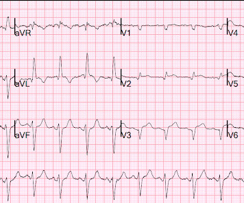
It turns out that the conventional algorithm was also worried, and because of that, the patient was brought to the criticalcare area. The STE in V1 is out of proportion to the S-wave, so V1 is also very worrisome (something I did not see on my phone). Probably significant infarction.
The software generates accurate reports in minutes using inexpensive devices, enabling emergency medicine, criticalcare, internal medicine, hospitalists and primary care physicians to make informed care decisions at the bedside. James Hillis, MBBS, DPhil, director of Clinical Operations at Mass General Brigham AI.
He was rushed by residents into our criticalcare room with a diagnosis of STEMI, and they handed me this ECG: There is sinus tachycardia with ST elevation in II, III, and aVF, as well as V4-V6. He presented to the Emergency Department with a blood pressure of 111/66 and a pulse of 117. He had this ECG recorded.
First troponin I returns at 48 ng/L ECG 5 143 min No significant change ECG 6 261 min Same hs Troponin I profile (peaked at 1849): Formal Echocardiogram SUMMARY The estimated left ventricular ejection fraction is 74 %. Eur Heart J 2018. Full text link. The estimated pulmonary artery systolic pressure is 27 mmHg + RA pressure.
Here is the post PCI EKG: And a few hours after that: The post PCI echocardiogram showed: Normal estimated left ventricular ejection fraction, 57%. Plus he did a 2 year combined EM Cardiology and CriticalCare Fellowship. Regional wall motion abnormality-mid to basal inferior wall.
This EKG was recorded as part of a standing order for criticalcare. After discussing all of the above with ED staff, we have made a decision to get stat echocardiogram and assess overall LV function and wall motion abnormalities and defer cath lab activation at the time." He had been smoking an opiate and suddenly collapsed.
Given the presentation, the cardiologist stented the vessel and the patient returned to the ICU for ongoing criticalcare. Echocardiogram showed LVEF 66% with normal wall motion and normal diastolic function. Lesions less than 70% are generally considered to be non-flow limiting. Two subsequent troponins were down trending.
Colin is an emergency medicine resident beginning his criticalcare fellowship in the summer with a strong interest in the role of ECG in criticalcare and OMI. Written by Colin Jenkins. Edits by Willy Frick. This confirms the suspicion of prior anterior OMI. The thrombus is circled below in red.
We organize all of the trending information in your field so you don't have to. Join thousands of users and stay up to date on the latest articles your peers are reading.
You know about us, now we want to get to know you!
Let's personalize your content
Let's get even more personalized
We recognize your account from another site in our network, please click 'Send Email' below to continue with verifying your account and setting a password.


Let's personalize your content