This site uses cookies to improve your experience. To help us insure we adhere to various privacy regulations, please select your country/region of residence. If you do not select a country, we will assume you are from the United States. Select your Cookie Settings or view our Privacy Policy and Terms of Use.
Cookie Settings
Cookies and similar technologies are used on this website for proper function of the website, for tracking performance analytics and for marketing purposes. We and some of our third-party providers may use cookie data for various purposes. Please review the cookie settings below and choose your preference.
Used for the proper function of the website
Used for monitoring website traffic and interactions
Cookie Settings
Cookies and similar technologies are used on this website for proper function of the website, for tracking performance analytics and for marketing purposes. We and some of our third-party providers may use cookie data for various purposes. Please review the cookie settings below and choose your preference.
Strictly Necessary: Used for the proper function of the website
Performance/Analytics: Used for monitoring website traffic and interactions
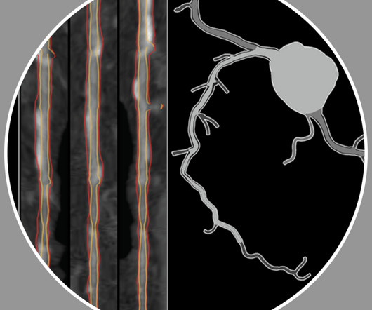
HeartFlow’s Plaque Analysis uses proprietary algorithms to analyze coronary CT angiogram (CCTA) scans, creating a personalized 3D model that quantifies and characterizes plaque volume in the coronaryarteries, aiding risk assessment of coronaryarterydisease. Narula et al. Rinehart et al. JSCAI 2024.
Reversing or regressing coronaryarterydisease is possible. But can coronaryarterydisease be reversed with lifestyle measures, including changes to nutrition and exercise? Subscribe now 1 Pathophysiology of CoronaryArteryDisease. In: Yuan, C., Hatsukami, T., Mossa-Basha, M.
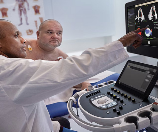
Image courtesy: Philips christine.book Wed, 06/12/2024 - 14:07 June 12, 2024 — Royal Philips has announced its next-generation AI-enabled cardiovascular ultrasound platform to help speed up cardiac ultrasound analysis with proven AI technology and reduce the burden on echocardiography labs.
Background Percutaneous coronary intervention (PCI) is a well-established treatment for coronaryarterydisease, one of the most significant causes of morbidity and mortality worldwide.
This study aims to investigate the relationship between sdLDLC level and PP in patients with stable coronaryartery disease.MethodsWe conducted a retrospective analysis of 146 lesions in 86 patients by repeat intravascular ultrasound examinations from January 2020 to May 2023.
Philips’ ultrasound AI strategy took another big step this week, with the launch of its next-generation echo AI platform, which will come integrated with the company’s cardiovascular ultrasound systems and bring a range of new echo-automating capabilities.
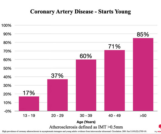
BackgroundStudies in young patients with stroke identified coronaryarterydisease (CAD) as a main contributor to mortality. In patients without previous CAD, but femoral plaque on ultrasound, nearly a half had nonobstructive and onefourth had obstructive CAD. Journal of the American Heart Association, Ahead of Print.
The algorithm uses deep learning to analyse routine ultrasound scans of the heart ( echocardiograms ) to detect disease that often goes undetected during standard assessments.
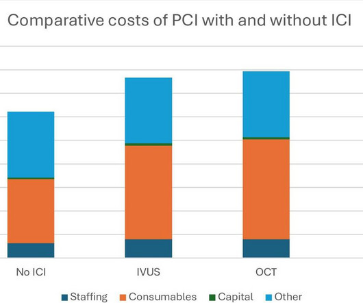
Stone, MD Mount Sinai Health System tim.hodson Wed, 04/02/2025 - 15:26 March 31, 2025 Using intravascular imaging (IVI) to guide stent implantation during complex stenting procedures is safer and more effective for patients with severely calcified coronaryarterydisease than conventional angiography, the more commonly used technique.
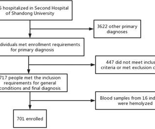
Background To investigate the correlation between lg (circSCMH1/miR-874) and acute coronary syndrome (ACS), acute myocardial infarction (AMI), and carotid plaque stability. Methods 701 patients were divided into stable coronaryarterydisease (SCAD), ACS, and control groups.
But the goal in this instance is to die after a long and healthy life ‘ with ’ coronaryarterydisease rather than ‘ from ’ coronaryarterydisease. However, the chances of dying from heart disease are directly proportional to the amount of plaque in your coronaryarteries.
Myocardial infarction with non-obstructive coronaryarteries (MINOCA) defines a heterogeneous group of atherosclerotic and non-atherosclerotic conditions, causing myocardial injury in the absence of obstructive coronaryarterydisease.
Background:Lipoprotein a (Lp(a)) is known to be associated with coronaryarterydisease and carotid artery atherosclerosis. Carotid ultrasound results were divided into two groups based on the presence or absence of plaque. Stroke, Volume 55, Issue Suppl_1 , Page ATP226-ATP226, February 1, 2024.
Patients usually have a normal life expectancy unless other structural heart diseases are present. An intravascular ultrasound was also performed, which was negative for vessel dissection. Introduction:Dextrocardia is a rare congenital condition where the heart's apex points to the right, with an incidence of about 0.01%.
More past history: hypertension, tobacco use, coronaryarterydisease with two vessel PCI to the right coronaryartery and circumflex artery several years prior. It is unknown when this pain recurred and became constant. He reports feeling nauseated with emesis. Then he was placed in a room after 30 minutes.
Angiogram No obstructive epicardial coronaryarterydisease Cannot exclude non-ACS causes of troponin elevation including coronary vasospasm, stress cardiomyopathy, microvascular disease, etc. Plaque rupture or erosion has been diagnosed by intravascular ultrasound in about 40 percent of women with MINOCA [ 12 ].
On arrival, lung ultrasound confirmed pulmonary edema (B lines). Mild Plaque no angiographically significant obstructive coronaryarterydisease. There is STE and hyperacute T-waves in V2 and V3, with significant STE in I and aVL, and inferior reciprocal STD. This is proximal LAD Occlusion until proven otherwise.
Aim The aim of this study was to determine the best clinical predictors of acute heart failure needing mechanical ventilation (MV) in the first 48 h of evolution of patients admitted because of acute coronary syndrome (ACS). Methods We analyzed a cohort of patients admitted for ACS between February 2017 and February 2018.
MINOCA: Myocardial Infarction in the Absence of Obstructive CoronaryArteryDisease). Here is my comment on MINOCA: "Non-obstructive coronarydisease" does not necessarily imply "no plaque rupture with thrombus." 2) overlooked obstructive coronarydisease (e.g., What is MINOCA? myocarditis).
This case was provided by Spencer Schwartz, an outstanding paramedic at Hennepin EMS who is on Hennepin EMS's specialized "P3" team, a team that receives extra training in advanced procedures such as RSI, thoracostomy, vasopressors, and prehospital ultrasound. Takotsubo is a sudden event, not one with crescendo angina. Lindahl et al.
Studies of patients with coronaryarterydisease who developed arrhythmic storm with episodes of PMVT following MI — show arrhythmias indistinguishable from those reported in this case. Smith comment: I agree with starting with a beta blocker such as esmolol, since this is likely to be a hyper-catecholaminergic state.
You can easily imagine this patient getting one of several diagnoses -- vasospasm, MINOCA , pericarditis, or maybe even no diagnosis at all beyond "non-obstructive coronaryarterydisease." Fortunately, this operator used intravascular ultrasound (IVUS). That plaque is at risk of thrombosing again.
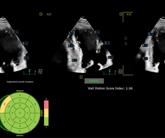
Given her risk factors (HTN, HLD, ESRD from diabetes) I decided to obtain a broad cardiac workup for the patient: serial ECGs, labs, serial troponins, CXR and bedside cardiac ultrasound. Ultrasounds can be very helpful in guiding your diagnostic pathway: location of WMA on US led to obtaining posterior leads.
No pericardial effusion on ultrasound." Cath lab activation by the ED and I agree with coronary angiography emergently." Result: no angiographically significant obstructive coronaryarterydisease. Healthy male under 25 years old with a pretty good story for acute onset crushing chest pain relieved with nitro.
Diffuse ST depression with ST elevation in aVR: Is this pattern specific for global ischemia due to left main coronaryarterydisease? Incidence of an acute coronary occlusion. Diffuse ST depression with ST elevation in aVR: Is this pattern specific for global ischemia due to left main coronaryarterydisease?
Smith comment: This patient did not have a bedside ultrasound. Had one been done, it would have shown a feature that is apparent on this ultrasound (however, this patient's LV function would not be as good as in this clip): This is recorded with the LV on the right. In fact, bedside ultrasound might even find severe aortic stenosis.
Echocardiography – We can use ultrasound to visualize the heart and look at how well it pumps. The best way to know if there is plaque in the heart arteries is by a test called CTCA (CT coronary angiography). This is termed as diastolic dysfunction. So what tests tell us about the heart as a pump?
The most common way to assess the presence and extent of coronaryarterydisease is with a CT scan, called a CT CAC score or CT Coronary Angiogram. CT Coronary Angiogram. 3 CORONARYDISEASE AMONG UNITED STATES SOLDIERS KILLED IN ACTION IN KOREA: PRELIMINARY REPORT. Circulation. Circulation.
However, the observed incongruity between carotid disease and the presence and severity of coronaryarterydisease (CAD) suggests differing mechanisms. Background:The presence of carotid plaque (CP) may serve as an indicator of panvascular atherosclerosis. Subsequently, patients were followed for 5.5
The clinical presentation of ischaemic heart disease has a broad spectrum, encompassing not only traditional epicardial coronaryarterydisease (CAD) characterised by stenosis or occlusion but also structural and functional disorders at both macrovessel and microvessel levels, as highlighted in the latest European guidelines.
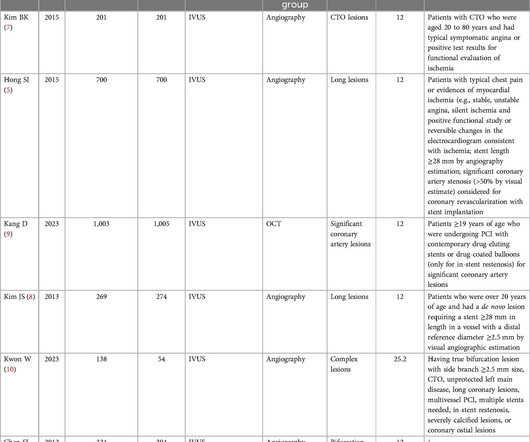
BackgroundThis study is to investigate the efficacy of stent implantation in patients with complex coronaryarterydisease (CAD) under intravascular ultrasound (IVUS) guidance and non-IVUS guidance.MethodsWe conducted a systematic search in PubMed, Web of Science, Cochran, and Embase for the articles of IVUS-guided and non-IVUS-guided stent implantation (..)
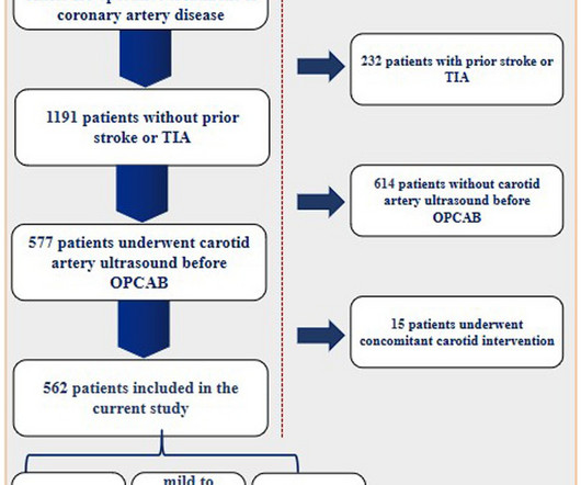
All enrolled patients underwent carotid arteryultrasound prior to OPCAB. The information was extracted independently by two authors of the study from the medical records.
I suspect pulmonary edema, but we are not given information on presence of B-lines on bedside ultrasound, or CXR findings. Anything that causes pulmonary edema: poor LV function, fluid overload, previous heart failure (HFrEF or HFpEF), valvular disease. Or I suspect that there is OMI simultaneous with another pathology.
We organize all of the trending information in your field so you don't have to. Join thousands of users and stay up to date on the latest articles your peers are reading.
You know about us, now we want to get to know you!
Let's personalize your content
Let's get even more personalized
We recognize your account from another site in our network, please click 'Send Email' below to continue with verifying your account and setting a password.


Let's personalize your content