This site uses cookies to improve your experience. To help us insure we adhere to various privacy regulations, please select your country/region of residence. If you do not select a country, we will assume you are from the United States. Select your Cookie Settings or view our Privacy Policy and Terms of Use.
Cookie Settings
Cookies and similar technologies are used on this website for proper function of the website, for tracking performance analytics and for marketing purposes. We and some of our third-party providers may use cookie data for various purposes. Please review the cookie settings below and choose your preference.
Used for the proper function of the website
Used for monitoring website traffic and interactions
Cookie Settings
Cookies and similar technologies are used on this website for proper function of the website, for tracking performance analytics and for marketing purposes. We and some of our third-party providers may use cookie data for various purposes. Please review the cookie settings below and choose your preference.
Strictly Necessary: Used for the proper function of the website
Performance/Analytics: Used for monitoring website traffic and interactions
Clinical introduction A woman in her 60s with non-obstructive coronaryarterydisease, aortic valve replacement and aortic arch repair, chronic diastolic heart failure and paroxysmal atrial fibrillation (AF) and flutter (AFL), presented with 3 days of sustained palpitations that felt similar to prior episodes of AF/AFL.
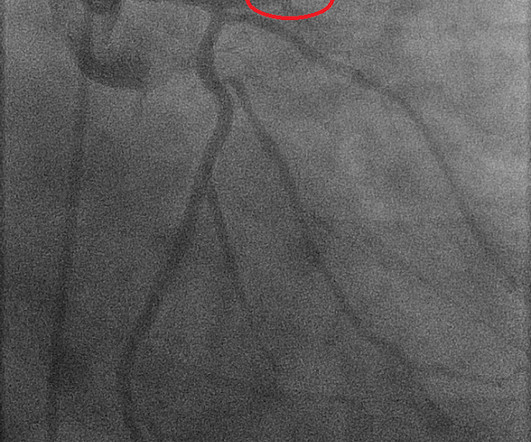
This ultimately resulted in severe coronaryartery occlusion, myocardial scarring and frequent episodes of ventricular tachycardia, which had a significant impact on the patient's quality of life. It is recommended that young and middle-aged patients with severe coronaryartery stenosis undergo screening for embolism.
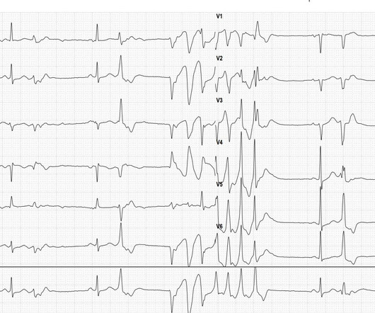
Then, a polymorphic ventricular tachycardia occurs over 7 beats. The QT interval of the sinus beats does not appear prolonged, thus ruling out Torsades de Pointes tachycardia. The most likely cause of this type of polymorphic ventricular tachycardia during a stress EKG is cardiac ischemia/coronaryarterydisease.
Introduction:Patients with Type 2 diabetes mellitus (T2DM) have an increased risk for coronaryarterydisease (CAD) compared to patients without T2DM. Ventricular arrhythmias (VA), such as ventricular fibrillation and ventricular tachycardia, are the major causes of mortality among patients with CAD.
Here is her ED ECG: Here is the ED physician's interpretation: IMPRESSION UNCERTAIN REGULAR RHYTHM, wide complex tachycardia, likely p-waves. LEFT BUNDLE BRANCH BLOCK [120+ ms QRS DURATION, 80+ ms Q/S IN V1/V2, 85+ ms R IN I/aVL/V5/V6] Comparison Summary: LBBB and tachycardia are new. This is clearly ventricular tachycardia.
Epicardial catheter ablation of ventricular tachycardia (VT) is a well-established ablation technique for a variety of myocardial substrates including ischemic cardiomyopathy. incidence of coronaryartery (CA) injury. Epicardial VT ablation has several potential life-threatening complications including a 1.5%
A 70-year-old male with a history of hypertension, hyperlipidemia, coronaryarterydisease, and CABG, presented with symptoms of moderate-intensity palpitations that terminated with Valsalva maneuvers. He was noted to have supraventricular tachycardia (SVT) on an event monitor.
A 72-year-old man with severe coronaryarterydisease (CAD) and coronaryartery bypass grafting complicated by ischemic cardiomyopathy and ventricular tachycardia (VT) was referred for redo VT ablation.
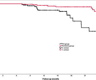
Regular clinical follow-ups were conducted to detect AF recurrence, defined as any episode of atrial fibrillation, atrial flutter or atrial tachycardia lasting >30 s. Results A total of 246 patients were enrolled, with 108 patients in the PVI group and 138 patients in the PVI+SVCI group.
There is increased LV cavity dimensions with an increase in transient ischemic dilation, suggesting Left Main, or 3-vessel coronaryarterydisease. 3. Sudden narrowing of a coronaryartery due to ACS (plaque rupture with thrombosis and/or downstream showering of platelet-fibrin aggregates). Type II ischemia.
This is ischemic ST depression, and could be due to increasing tachycardia, with a heart rate over 130, but that is unlikely given that the patient is now complaining of crushing chest pain and that there was tachycardia all along. One would expect that the angiogram would show open arteries with normal TIMI-3 flow and culprit lesions.
ECG in a person with persistent anginal pain for the past several hours showing significant ST segment depression anterolateral leads along with sinus tachycardia. Such a pattern is consistent with significant left main coronaryartery stenosis. ST elevation in aVR more than that in V1 is also suggestive of left main disease.
Sometimes, head up tilt test, also known in short as HUTT, is also done for the evaluation of postural orthostatic tachycardia syndrome, POTS, a condition in which there is tachycardia on standing up, without a fall in blood pressure. Transcript of the video: Head up tilt test, is usually done for the evaluation of recurrent syncope.
I quickly reviewed the patient’s records and saw that she was a 53 year old woman with a history of BMI 40, but no other identifiable risk factors for coronaryarterydisease. The absence of risk factors for coronaryarterydisease does not mean a patient is not at risk for OMI.
He was counseled to abstain from cannabis use.Conclusion:At low to moderate doses, cannabis can lead to a surge in sympathetic activity causing tachycardia and hypertension, while parasympathetic activity is predominant at higher doses, causing bradycardia and hypotension. Patient did not report any symptoms and was hemodynamically stable.
A 56 year old male with a history of diabetes, dyslipidemia, hypertension, and coronaryarterydisease presented to the emergency department with sudden onset weakness, fatigue, lethargy, and confusion. The conventional computer algorithm called “ sinus tachycardia, otherwise normal EKG ”.
Conventional algorithm interpretation: SINUS TACHYCARDIA ABNORMAL RHYTHM ECG Confirmed by over-reading physician Transformed ECG by PM Cardio: PM Cardio interpretation: OMI with Low Confidence Dr. Rob Reardon did a bedside echo using Speckle tracking. Mild Plaque no angiographically significant obstructive coronaryarterydisease.
Similarly, you may use our , app to adjust the paper speed along with amplification to read the slightest changes, especially for conditions like tachycardia and bradycardia. Alternatively, it also helps enhance arrhythmia management with coronaryarterydisease.
Diffuse ST depression with ST elevation in aVR: Is this pattern specific for global ischemia due to left main coronaryarterydisease? Incidence of an acute coronary occlusion. Diffuse ST depression with ST elevation in aVR: Is this pattern specific for global ischemia due to left main coronaryarterydisease?
Old ‘NSTEMI’ A history of coronaryarterydisease and a stent to the same territory further increases pre-test likelihood of acute coronary occlusion, including in-stent thrombosis. This ECG has Q-waves, but they are not very wide nor very deep, and so I doubt that the inferior STE is due to old MI.
He denied any known medical history, specifically: coronaryarterydisease, hypertension, dyslipidemia, diabetes, heart failure, myocardial infarction, or any prior PCI/stent. Vital signs were noted to be unremarkable with respect to any hypo-hypertensive crisis, hypoxia, etc. Breath sounds were clear in all lung fields.
A coronary angiogram was done that did not show significant coronaryarterydisease. But there are 3 other wide beats in the tachycardia that begins with beat #6 ( = beats #7; 13,14 ). Post ROSC the patient was alert and cooperative. Echocardiography showed apical ballooning with hypokinesis.
Angiogram --Minimal coronary atherosclerosis --No obstructive epicardial coronaryarterydisease or evidence of plaque rupture noted to explain prolonged QT or ventricular fibrillation cardiacarrest, suspect nonischemic mechanism Echo The estimated left ventricular ejection fraction is 45 %.
He was taken emergently to the cardiac catheterization lab and found to have multi-vessel coronaryarterydisease with a near-occlusive culprit lesion in the RCA, possibly reperfused. Slow TIMI 2 initially with brisk flow status post percutaneous coronary intervention with 18mm drug-eluting stent.
A CT Coronary angiogram was ordered. Here are the results: --Minimally obstructive coronaryarterydisease. --LAD That said, against acute PE — is the inconstancy of this patient's symptoms — the lack of tachycardia — and the lack of any other ECG signs of acute RV strain. A repeat troponin returned at 0.45
Diffuse ST depression with ST elevation in aVR: Is this pattern specific for global ischemia due to left main coronaryarterydisease? My L IST includes the following: i ) LVH with strain; ii ) Ischemia; iii ) Digoxin use; iv ) HypoKalemia and/or HypoMagnesemia; v ) Tachycardia; and , vi ) Any combination of i-thru-v.
The diagnostic coronary angiogram identified only minimal coronaryarterydisease, but there was a severely calcified, ‘immobile’ aortic valve. 3) Anemia, or poisons of hemoglobin such as methemoglobin or CO 4) Fixed coronary stenosis that limits flow. In the cath lab, the patient’s blood pressure remained low.
We identified the subset of variables most predictive of post‐HSCT cardiovascular events, defined as a composite of cardiovascular death, myocardial infarction, heart failure, stroke, atrial fibrillation or flutter, and sustained ventricular tachycardia.
An external monitor revealed one episode of non-sustained supraventricular tachycardia, otherwise was unremarkable. Cardiac catheterization revealed non-obstructive coronaryarterydisease. He experienced worsening symptoms for which emergency department evaluation revealed anterior lead T-wave inversions on EKG.
The ST segment changes are compatible with severe subendocardial ischemia which can be caused by type I MI from ACS or potentially from type II MI (non-obstructive coronaryarterydisease with supply/demand mismatch).
A fast heartbeat is called tachycardia, while a slow heartbeat is called bradycardia in medical terms. Coronaryarterydisease Excessive cholesterol builds up plaque that blocks the arteries supplying blood to the heart. ECG machine can also help diagnose coronaryarterydisease.
This progressed to electrical storm , with incessant PolyMorphic Ventricular Tachycardia ( PMVT ) and recurrent episodes of Ventricular Fibrillation ( VFib ). Studies of patients with coronaryarterydisease who developed arrhythmic storm with episodes of PMVT following MI — show arrhythmias indistinguishable from those reported in this case.
Here is another proven left main occlusion in a young woman who presented with sudden pulmonary edema, had this ECG recorded, then arrested and was resuscitated after 30 minutes of CPR: This has sinus tachycardia with RBBB and LAFB, and STE in V2-V6 as well as I, aVL This pattern could just as easily be seen in LAD occlusion.
It’s essential for those at risk of coronaryarterydisease to be aware of the following symptoms. The abnormal heart rhythms can further lead to death because of ventricular tachycardia and ventricular fibrillation. High triglyceride levels may also increase your risk of coronaryarterydisease, particularly in women.
Written by Willy Frick with edits by Ken Grauer An older man with a history of non-ischemic HFrEF s/p CRT and mild coronaryarterydisease presented with chest pain. Additionally, the patient had no other apparent reason to have sinus tachycardia (such as volume depletion, bleeding, fever). ECG 1 What do you think?
The status of the patients chest pain at this time is unknown : EKG 1, 1300: There is sinus tachycardia and artifact of low and high frequency. However, there is also significant tachycardia , with heart rate of 116, and known hypoxia. The scan showed a bicuspid aortic valve with severe stenosis and coronaryarterydisease.
September 2024 Approvals Flurpiridaz F 18 Approval Date: September 27, 2024 Indication: A radioactive positron emission tomography (PET) tracer for imaging myocardial ischemia and infarction in adults with suspected or known coronaryarterydisease. Studies on Supraventricular Tachycardia : 5 studies , a total of 317 adults.
Ventricular tachycardia?) He said it felt like "someone ripped [his] heart out." While walking into the ED, the patient had an episode of "dizziness" causing him to fall onto his stomach. What do you think? I sent this ECG to Dr. Smith and Dr. Meyers with no context. Dr. Smith immediately replied "Swirl. Proximal LAD." Dr. Meyers agreed.
Written by Pendell Meyers A woman in her 70s with known prior coronaryarterydisease experienced acute chest pain and shortness of breath. KEY Points: DSI does not indicate acute coronary occlusion! This pattern of DSI does not mean there is acute coronary occlusion. Vital signs were within normal limits.
Circumstances attending 100 sudden deaths from coronaryarterydisease with coroners necropsies. This was overtaken by a predominance of sympathetic surge ( tachycardia, persistent ST elevation development of electrical "storm" with failure to respond to recurrent defibrillation ). Circulation , 125 (3), 491496.
We organize all of the trending information in your field so you don't have to. Join thousands of users and stay up to date on the latest articles your peers are reading.
You know about us, now we want to get to know you!
Let's personalize your content
Let's get even more personalized
We recognize your account from another site in our network, please click 'Send Email' below to continue with verifying your account and setting a password.


Let's personalize your content