This site uses cookies to improve your experience. To help us insure we adhere to various privacy regulations, please select your country/region of residence. If you do not select a country, we will assume you are from the United States. Select your Cookie Settings or view our Privacy Policy and Terms of Use.
Cookie Settings
Cookies and similar technologies are used on this website for proper function of the website, for tracking performance analytics and for marketing purposes. We and some of our third-party providers may use cookie data for various purposes. Please review the cookie settings below and choose your preference.
Used for the proper function of the website
Used for monitoring website traffic and interactions
Cookie Settings
Cookies and similar technologies are used on this website for proper function of the website, for tracking performance analytics and for marketing purposes. We and some of our third-party providers may use cookie data for various purposes. Please review the cookie settings below and choose your preference.
Strictly Necessary: Used for the proper function of the website
Performance/Analytics: Used for monitoring website traffic and interactions
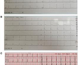
The principal clinical manifestation of thrombophilia is venous thromboembolism, which is also markedly linked to arterialthrombosis, including myocardial infarction. The patient had a history of deep vein thrombosis and was genetically tested to carry two thrombophilia susceptibility alleles at the PAI-1 (4G/5G) and MTHFR (C>T) loci.
Background Kounis syndrome is an acute coronary syndrome (ACS) caused by allergic reactions, including coronaryartery spasm (type I) caused by allergies without coronary predisposing factors, pre-existing coronary atherosclerosis, and coronaryarterydisease.
There is increased LV cavity dimensions with an increase in transient ischemic dilation, suggesting Left Main, or 3-vessel coronaryarterydisease. 3. Coronary angiography reveals significant and severe CAD involving all three epicardial vessels. A large Diagonal artery has subtotal occlusion proximally.
Angiogram No obstructive epicardial coronaryarterydisease Cannot exclude non-ACS causes of troponin elevation including coronary vasospasm, stress cardiomyopathy, microvascular disease, etc. The degree of stenosis is not a great predictor of thrombosis, and culprits may not be visible.
Hospital Course The patient was taken emergently to the cath lab which did not reveal any significant coronaryarterydisease, but she was noted to have reduced EF consistent with Takotsubo cardiomyopathy. An apical OMI has the same ultrasound findings as takotsubo, and thus mimics takotsubo. Learning Points: 1.
Coronary angiography revealed a tortuous and extremely aneurysmal RCA, as well as multivessel coronaryarterydisease (mvCAD) involving LAD, D1, LCx, OM1. Notably, the LAD had multiple aneurysmal segments and areas of eccentric stenosis upto 90%.Multislice
Old ‘NSTEMI’ A history of coronaryarterydisease and a stent to the same territory further increases pre-test likelihood of acute coronary occlusion, including in-stent thrombosis. So this NSTEMI was likely a STEMI(-)OMI with delayed reperfusion.
History sounds concerning for ACS (could be critical stenosis, triple vessel), but differential also includes dissection, GI bleed, etc. Diffuse ST depression with ST elevation in aVR: Is this pattern specific for global ischemia due to left main coronaryarterydisease? Incidence of an acute coronary occlusion.
MINOCA: Myocardial Infarction in the Absence of Obstructive CoronaryArteryDisease). Here is my comment on MINOCA: "Non-obstructive coronarydisease" does not necessarily imply "no plaque rupture with thrombus." 2) overlooked obstructive coronarydisease (e.g., What is MINOCA? myocarditis).
The cardiologist called this 20% stenosis. You can easily imagine this patient getting one of several diagnoses -- vasospasm, MINOCA , pericarditis, or maybe even no diagnosis at all beyond "non-obstructive coronaryarterydisease." Smith comment : a very high proportion of MINOCA are ruptured plaque with lysed thrombus.
Coronary angiography gives a visual impression about the severity of the stenosis. But it need not imply the actual functional significance of the stenosis in terms of flow physiology. FAME study showed that at one year follow up, rate of major adverse coronary events was reduced by approximately 30% by routinely measuring FFR.
Background Untreated multivessel disease (MVD) in acute myocardial infarction (AMI) has been linked to a higher risk of recurrent ischemia and death within one year. Similarly, all-cause mortality, cardiovascular mortality, stent thrombosis, and acute renal insufficiency did not show significant differences between two groups.
SMART 4 ( NCT04722250 ) studied patients with severe aortic stenosis and a small aortic annulus who underwent transcatheter aortic valve replacement (TAVR). The primary non-inferiority endpoint was MACCE (a composite of cardiac death, MI, ischaemic stroke, stent thrombosis, or target vessel revascularisation).
Decedents with acute coronarythrombosis, myocardial infarction, or other myocardial abnormality were excluded. Decedents with either noncardiac death or SAD had similar height, weight, and heart weight. Moreover, decedents with SAD had lower cardiomyocyte width (mean, 18.6 m versus 19.6 m; mean difference, 1.0 m [95% CI, 0.21.8],P=0.014)
We organize all of the trending information in your field so you don't have to. Join thousands of users and stay up to date on the latest articles your peers are reading.
You know about us, now we want to get to know you!
Let's personalize your content
Let's get even more personalized
We recognize your account from another site in our network, please click 'Send Email' below to continue with verifying your account and setting a password.


Let's personalize your content