This site uses cookies to improve your experience. To help us insure we adhere to various privacy regulations, please select your country/region of residence. If you do not select a country, we will assume you are from the United States. Select your Cookie Settings or view our Privacy Policy and Terms of Use.
Cookie Settings
Cookies and similar technologies are used on this website for proper function of the website, for tracking performance analytics and for marketing purposes. We and some of our third-party providers may use cookie data for various purposes. Please review the cookie settings below and choose your preference.
Used for the proper function of the website
Used for monitoring website traffic and interactions
Cookie Settings
Cookies and similar technologies are used on this website for proper function of the website, for tracking performance analytics and for marketing purposes. We and some of our third-party providers may use cookie data for various purposes. Please review the cookie settings below and choose your preference.
Strictly Necessary: Used for the proper function of the website
Performance/Analytics: Used for monitoring website traffic and interactions
Old ‘NSTEMI’ A history of coronaryarterydisease and a stent to the same territory further increases pre-test likelihood of acute coronary occlusion, including in-stent thrombosis. So this NSTEMI was likely a STEMI(-)OMI with delayed reperfusion. Deutch et al.
This is ischemic ST depression, and could be due to increasing tachycardia, with a heart rate over 130, but that is unlikely given that the patient is now complaining of crushing chest pain and that there was tachycardia all along. One would expect that the angiogram would show open arteries with normal TIMI-3 flow and culprit lesions.
This has been termed a “STEMI equivalent” and included in STEMI guidelines, suggesting this patient should receive dual anti-platelets, heparin and immediate cath lab activation–or thrombolysis in centres where cath lab is not available. aVR ST segment elevation: acute STEMI or not? Incidence of an acute coronary occlusion.
A 56 year old male with a history of diabetes, dyslipidemia, hypertension, and coronaryarterydisease presented to the emergency department with sudden onset weakness, fatigue, lethargy, and confusion. At 2111, the troponin I peaked at 12.252 ng/mL (this is in the range of STEMI patients, quite high).
Prehospital Conventional algorithm interpretation: ANTERIOR INFARCT, STEMI Transformed ECG by PM Cardio: PM Cardio AI Bot interpretation: OMI with High Confidence What do you think? Mild Plaque no angiographically significant obstructive coronaryarterydisease. She had acute pulmonary edema on exam.
He denied any known medical history, specifically: coronaryarterydisease, hypertension, dyslipidemia, diabetes, heart failure, myocardial infarction, or any prior PCI/stent. It doesn’t meet any conventional STEMI criteria, but there is patently obvious increased area under the curve. No appreciable skin pallor.
A CT Coronary angiogram was ordered. Here are the results: --Minimally obstructive coronaryarterydisease. --LAD Although a lesion is not visible anatomically on this CT scan, coronary catheter angiography could be considered based on Cardiology evaluation." It is likely that the artery will re-occlude.
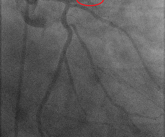
He was taken emergently to the cardiac catheterization lab and found to have multi-vessel coronaryarterydisease with a near-occlusive culprit lesion in the RCA, possibly reperfused. Slow TIMI 2 initially with brisk flow status post percutaneous coronary intervention with 18mm drug-eluting stent.
Clinical Course The paramedic activated a “Code STEMI” alert and transported the patient nearly 50 miles to the closest tertiary medical center. The diagnostic coronary angiogram identified only minimal coronaryarterydisease, but there was a severely calcified, ‘immobile’ aortic valve. What do you see?
Diffuse ST depression with ST elevation in aVR: Is this pattern specific for global ischemia due to left main coronaryarterydisease? My L IST includes the following: i ) LVH with strain; ii ) Ischemia; iii ) Digoxin use; iv ) HypoKalemia and/or HypoMagnesemia; v ) Tachycardia; and , vi ) Any combination of i-thru-v.
The ECG shows obvious STEMI(+) OMI due to probable proximal LAD occlusion. This progressed to electrical storm , with incessant PolyMorphic Ventricular Tachycardia ( PMVT ) and recurrent episodes of Ventricular Fibrillation ( VFib ). The below ECG was recorded. He required multiple defibrillations within a period of a few hours.
When total LM occlusion does present with STE in aVR, there is ALWAYS ST Elevation elsewhere which makes STEMI obvious; in other words, STE is never limited to only aVR but instead it is part of a massive and usually obvious STEMI. All are, however, clearly massive STEMI. This is her ECG: An obvious STEMI, but which artery?
The status of the patients chest pain at this time is unknown : EKG 1, 1300: There is sinus tachycardia and artifact of low and high frequency. However, there is also significant tachycardia , with heart rate of 116, and known hypoxia. Supply-demand mismatch can cause ST Elevation (Type 2 STEMI). An EKG was immediately recorded.
Ventricular tachycardia?) Learning points: 40% of LAD OMI with TIMI-0 flow do NOT meet STEMI criteria (manuscript under consideration at Eur Ht. 20/53 did not meet STEMI criteria, but all showed OMI diagnosed by both Smith and the Queen of Hearts. He said it felt like "someone ripped [his] heart out." What do you think?
Written by Pendell Meyers A woman in her 70s with known prior coronaryarterydisease experienced acute chest pain and shortness of breath. Final Diagnosis: "STEMI" (of course, as you can see in the ECGs above, this is not true, by definition this was NSTEMI. KEY Points: DSI does not indicate acute coronary occlusion!
Circumstances attending 100 sudden deaths from coronaryarterydisease with coroners necropsies. Frick the ongoing ECG changes in association with hemodynamic collapse seemed out of proportion to the apparent coronary pathology. Risk of acute myocardial infarction after the death of a significant person in ones life.
We organize all of the trending information in your field so you don't have to. Join thousands of users and stay up to date on the latest articles your peers are reading.
You know about us, now we want to get to know you!
Let's personalize your content
Let's get even more personalized
We recognize your account from another site in our network, please click 'Send Email' below to continue with verifying your account and setting a password.


Let's personalize your content