This site uses cookies to improve your experience. To help us insure we adhere to various privacy regulations, please select your country/region of residence. If you do not select a country, we will assume you are from the United States. Select your Cookie Settings or view our Privacy Policy and Terms of Use.
Cookie Settings
Cookies and similar technologies are used on this website for proper function of the website, for tracking performance analytics and for marketing purposes. We and some of our third-party providers may use cookie data for various purposes. Please review the cookie settings below and choose your preference.
Used for the proper function of the website
Used for monitoring website traffic and interactions
Cookie Settings
Cookies and similar technologies are used on this website for proper function of the website, for tracking performance analytics and for marketing purposes. We and some of our third-party providers may use cookie data for various purposes. Please review the cookie settings below and choose your preference.
Strictly Necessary: Used for the proper function of the website
Performance/Analytics: Used for monitoring website traffic and interactions
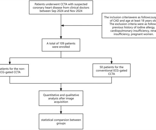
ObjectiveTo compare the image quality, radiation dose, and examination time between non-electrocardiogram (ECG)-gated coronary CT angiography (ECG-less CCTA) and conventional ECG-gated CCTA using wide-detector CT, and validate its clinical applicability.MethodsIn this prospective study, 109 patients with suspected coronaryarterydisease were divided (..)
Coronaryarterydisease (CAD) is a leading cause of mortality worldwide. It slows ventricular conduction and increases dispersion of repolarization, manifesting differently on the electrocardiogram (ECG).
Data on the prognostic significance of temporal variability of spatial heterogeneity of electrocardiographic repolarization in coronaryarterydisease (CAD) are limited.
Background Kounis syndrome is an acute coronary syndrome (ACS) caused by allergic reactions, including coronaryartery spasm (type I) caused by allergies without coronary predisposing factors, pre-existing coronary atherosclerosis, and coronaryarterydisease. ng/ml(0–0.5 ng/ml).
Objective Cardiac cephalalgia, once seen as a rare symptom of coronaryarterydisease, is now more recognized. His headache improved after percutaneous coronary intervention. Electrocardiogram (ECG) might not always show abnormalities, and chest pain is not always present.
Patients usually have a normal life expectancy unless other structural heart diseases are present. Electrocardiogram (ECG) showed a prominent S wave in the left-sided leads and a prominent R wave in the right-sided chest leads, suggesting dextrocardia. His vital signs were normal, and the physical examination was unremarkable.
This highlights the need for serial ECGs in acute coronary syndrome as initial ECGs may be near normal even in those with severe disease. Classical electrocardiographic pattern in left main coronaryarterydisease is ST elevation in aVR with extensive ST depression in other leads, most prominent in I, II and V4-V6.
Electrocardiogram (ECG) and telemetry revealed junctional bradycardia with heart rate in 30s and sinus pauses (5-7 seconds). Patient did not report any symptoms and was hemodynamically stable. He was euvolemic on physical exam.
Diffuse ST depression with ST elevation in aVR: Is this pattern specific for global ischemia due to left main coronaryarterydisease? Incidence of an acute coronary occlusion. New insights into the use of the 12-lead electrocardiogram for diagnosing acute myocardial infarction in the emergency department.
You will note that it is essentially an unremarkable electrocardiogram except for some PACS. He was taken emergently to the cardiac catheterization lab and found to have multi-vessel coronaryarterydisease with a near-occlusive culprit lesion in the RCA, possibly reperfused.
In a study published in Communications Medicine , David Ouyang, MD, assistant professor of Cardiology and Medicine at Cedars-Sinai, along with Chugh and fellow investigators trained a deep learning algorithm to study patterns in electrocardiograms, also known as ECGs, which are recordings of the heart’s electrical activity.
Heart Valve Disease If one or more heart valves are not functioning correctly, it can cause blood to flow backward, putting extra pressure on the heart, which may cause it to expand to compensate for the inefficiency. This may result in ischemia (lack of oxygen to the heart muscle), causing parts of the heart to weaken and enlarge.
Background Patients with low HEART (History, Electrocardiogram, Age, Risk factors, and Troponin level) risk scores who are discharged from the emergency department (ED) may present clinical challenges and diagnostic dilemmas. The use of downstream non-invasive stress imaging (NISI) tests in this population remains uncertain.
An electrocardiogram is a machine used to record the heart's electrical activity. Electrocardiogram, echocardiogram, and some other tests are done for patients with cardiac arrest. Coronaryarterydisease Excessive cholesterol builds up plaque that blocks the arteries supplying blood to the heart.
D) An electrocardiogram is most commonly normal in these patients. Takotsubo Cardiomyopathy is distinguished by overall systolic dysfunction of the left ventricle (LV), replicating that of a myocardial infarction (heart attack), but without angiographic evidence of coronaryarterydisease or a blockage.
These tests may include: Electrocardiogram (ECG) : Records the electrical activity of your heart. Regular heart check-ups offer several benefits including: Early detection of heart disease : Many heart conditions such as high blood pressure, high cholesterol and coronaryarterydisease can be detected early through regular screenings.
LR was based on normal examination, stable hemodynamics, normal electrocardiograms (ECG), and negative cardiac troponin I, without pre-discharge functional or anatomic cardiac testing or risk scores. Length of stay (LOS) in the CPU to discharge was 10.4
Explanation: Shown electrocardiogram suggests left ventricular hypertrophy. Shown electrocardiogram suggests left ventricular hypertrophy. Start aspirin and Plavix Correct answer: (B) (B) Echocardiogram is indicated. Hypertrophic cardiomyopathy is one of them.
At the bottom of the post, I have re-printed the section on aVR in my article on the ECG in ACS from the Canadian Journal of Cardiology: New Insights Into the Use of the 12-Lead Electrocardiogram for Diagnosing Acute Myocardial Infarction in the Emergency Department Case 1. Updates on the Electrocardiogram in Acute Coronary Syndromes.
Integrating ECG Testing to identify cardiometabolic changes and measure the risk of CVD Electrocardiogram Machines like , Wellnest 12L Pro2 serve as a valuable tools in this endeavor, offering a window into the intricate relationship between ART and cardiovascular health.
Early Continuous ST Segment Monitoring in Unstable Angina: Prognostic Value Additional to the Clinical Characteristics and the Admission Electrocardiogram. Compared with sustained STEMI, transient STEMI was associated with less myocardial damage, less extensive coronaryarterydisease, higher TIMI flow grade, and better cardiac function.
He had undergone coronaryartery bypass grafting due to myocardial infarction and severe three-vessel coronaryarterydisease. Figure 1 (A) Resting 12-lead electrocardiogram. (B) B) Standard Limb II-lead electrocardiogram of Holter monitor. s (blue arrows; figure 1b ).
BackgroundAcute myocardial infarction commonly occurs in patients with coronaryarterydisease, but rarely, it can develop under a hypercoagulable state. These thrombotic complications predominantly arise in veins rather than in arteries.
BackgroundPainful left bundle branch block (LBBB) syndrome is an uncommon disease that is defined as intermittent episodes of angina associated with simultaneous LBBB changes on an electrocardiogram (ECG) with the absence of flow-limiting coronaryarterydisease or ischemia on functional testing.
Some murmurs caused by heart disease or coronaryarterydisease can improve with lifestyle changes, medication, or surgical procedures. Electrocardiogram (ECG): An ECG records the electrical activity of the heart and can help detect arrhythmias or other heart problems.
We organize all of the trending information in your field so you don't have to. Join thousands of users and stay up to date on the latest articles your peers are reading.
You know about us, now we want to get to know you!
Let's personalize your content
Let's get even more personalized
We recognize your account from another site in our network, please click 'Send Email' below to continue with verifying your account and setting a password.


Let's personalize your content