This site uses cookies to improve your experience. To help us insure we adhere to various privacy regulations, please select your country/region of residence. If you do not select a country, we will assume you are from the United States. Select your Cookie Settings or view our Privacy Policy and Terms of Use.
Cookie Settings
Cookies and similar technologies are used on this website for proper function of the website, for tracking performance analytics and for marketing purposes. We and some of our third-party providers may use cookie data for various purposes. Please review the cookie settings below and choose your preference.
Used for the proper function of the website
Used for monitoring website traffic and interactions
Cookie Settings
Cookies and similar technologies are used on this website for proper function of the website, for tracking performance analytics and for marketing purposes. We and some of our third-party providers may use cookie data for various purposes. Please review the cookie settings below and choose your preference.
Strictly Necessary: Used for the proper function of the website
Performance/Analytics: Used for monitoring website traffic and interactions
In this week’s View, Dr. Eagle looks at the difference between quantitative coronary angiography versus intervascular ultrasound to guide PCI. He then discusses paclitaxel-coated balloon catheters vs uncoated balloon angioplasty for treating coronary in-stent restenosis.
However, the patient's cardiac Doppler ultrasound indicated poor cardiac contractions, and extracorporeal membrane oxygenation (ECMO) was started immediately. Coronaryarterybypassgrafting was performed after the patient's condition stabilized, and he was finally discharged.
Smith comment: This patient did not have a bedside ultrasound. Had one been done, it would have shown a feature that is apparent on this ultrasound (however, this patient's LV function would not be as good as in this clip): This is recorded with the LV on the right. In fact, bedside ultrasound might even find severe aortic stenosis.
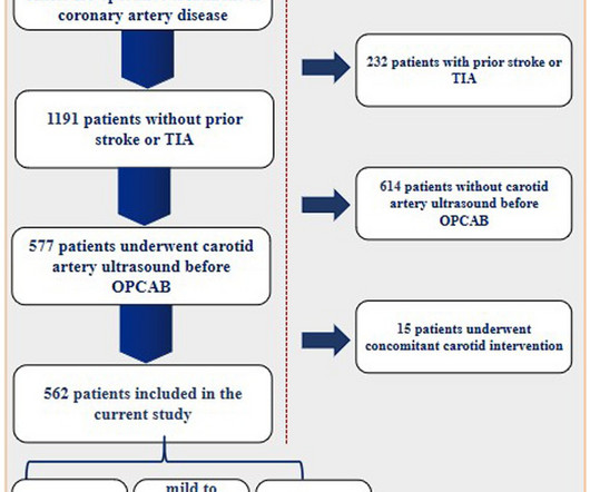
All enrolled patients underwent carotid arteryultrasound prior to OPCAB. The information was extracted independently by two authors of the study from the medical records. Both univariate and multivariate analyses were conducted.ResultsA total of 562 patients met the inclusion criteria for the current study.
IntroductionNumerous techniques have been developed to minimize risk of perioperative stroke during coronaryarterybypassgrafting (CABG), including off-pump approach, preoperative and intraoperative imaging of the ascending aorta (CT scan and epiaortic ultrasound), anaortic CABG with bilateral internal thoracic artery, clampless devices for the construction (..)
I suspect pulmonary edema, but we are not given information on presence of B-lines on bedside ultrasound, or CXR findings. The patient was again sent home with a plan for surgical aortic valve replacement and coronaryarterybypassgrafting to the PDA. We certainly know that there is hypoxia.
We organize all of the trending information in your field so you don't have to. Join thousands of users and stay up to date on the latest articles your peers are reading.
You know about us, now we want to get to know you!
Let's personalize your content
Let's get even more personalized
We recognize your account from another site in our network, please click 'Send Email' below to continue with verifying your account and setting a password.


Let's personalize your content