This site uses cookies to improve your experience. To help us insure we adhere to various privacy regulations, please select your country/region of residence. If you do not select a country, we will assume you are from the United States. Select your Cookie Settings or view our Privacy Policy and Terms of Use.
Cookie Settings
Cookies and similar technologies are used on this website for proper function of the website, for tracking performance analytics and for marketing purposes. We and some of our third-party providers may use cookie data for various purposes. Please review the cookie settings below and choose your preference.
Used for the proper function of the website
Used for monitoring website traffic and interactions
Cookie Settings
Cookies and similar technologies are used on this website for proper function of the website, for tracking performance analytics and for marketing purposes. We and some of our third-party providers may use cookie data for various purposes. Please review the cookie settings below and choose your preference.
Strictly Necessary: Used for the proper function of the website
Performance/Analytics: Used for monitoring website traffic and interactions
Coronaryarterybypassgrafting (CABG) is a surgical procedure that improves blood flow to the heart tissue and can effectively treat myocardial ischemia caused by coronaryarterydisease.
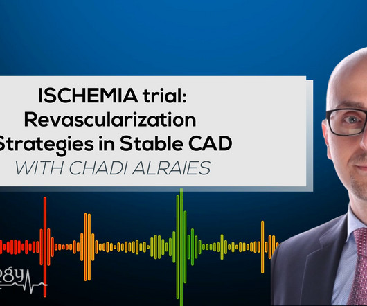
In the ISCHEMIA (International Study of Comparative Health Effectiveness with Medical and Invasive Approaches) trial, researchers examined the risk of ischemic events in patients with stable coronaryarterydisease. years, with 57.1% occurring within 30 days after CABG. Original article: Redfors B et al.
Angiography done after initial stabilization showed severe stenosis of distal left main coronaryartery. In addition, there were multiple lesions in all three vessels, making a standard indication for an urgent coronaryarterybypassgrafting. There is minimal ST segment elevation in aVR as well.
The patient was brought directly to the cardiac catheterization lab for PCI, bypassing the ED. The diagnostic coronary angiogram identified only minimal coronaryarterydisease, but there was a severely calcified, ‘immobile’ aortic valve. In the cath lab, the patient’s blood pressure remained low.
In most cases, rather, the culprit is gross ischemia due to myocardial infarction, cardiomyopathy, or advanced coronaryarterydisease. Unfortunately, today’s case is lacking any such diagnostics, thus I cannot say with certainty that the QT interval is, or is not, culpable in arrhythmogenesis. [1]
Watch what happends as the heart recovers from its episode of ischemia. Angiogram: Severe two-vessel coronaryarterydisease of a left dominant system including 70 to 80% stenosis involving the distal left main/bifurcation. The ECG shows inferior ischemia. Are the T-waves in leads I and II hyperacute? Hard to tell.
The patient was started on heparin for possible NSTEMI vs demand ischemia. increasing stenosis, ischemia, volume changes, increased blood pressure, atrial fibrillation, etc.) The scan showed a bicuspid aortic valve with severe stenosis and coronaryarterydisease. What "initiates" the aortic stenosis cascade?
We organize all of the trending information in your field so you don't have to. Join thousands of users and stay up to date on the latest articles your peers are reading.
You know about us, now we want to get to know you!
Let's personalize your content
Let's get even more personalized
We recognize your account from another site in our network, please click 'Send Email' below to continue with verifying your account and setting a password.


Let's personalize your content