This site uses cookies to improve your experience. To help us insure we adhere to various privacy regulations, please select your country/region of residence. If you do not select a country, we will assume you are from the United States. Select your Cookie Settings or view our Privacy Policy and Terms of Use.
Cookie Settings
Cookies and similar technologies are used on this website for proper function of the website, for tracking performance analytics and for marketing purposes. We and some of our third-party providers may use cookie data for various purposes. Please review the cookie settings below and choose your preference.
Used for the proper function of the website
Used for monitoring website traffic and interactions
Cookie Settings
Cookies and similar technologies are used on this website for proper function of the website, for tracking performance analytics and for marketing purposes. We and some of our third-party providers may use cookie data for various purposes. Please review the cookie settings below and choose your preference.
Strictly Necessary: Used for the proper function of the website
Performance/Analytics: Used for monitoring website traffic and interactions
Post cath ECG: Now there are hyperacute T-waves again, and recurrent ST depression in V2 This ECG would normally diagnostic of OMI until proven otherwise No further troponins were measured, but it looks like there is recurrent OMI Next day: A CT CoronaryAngiogram was done (CTCA) CARDIAC MORPHOLOGY AND FUNCTION: 1. IMPRESSION: 1.
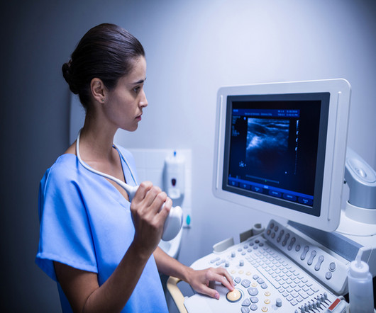
Bedside cardiac ultrasound showed moderately decreased LV function. Finally, do a coronaryangiogram Possible alternative to pacing is to give a beta-1 agonist to increase heart rate. She was intubated. CT of the chest showed no pulmonary embolism but bibasilar infiltrates. Dobutamine is an acceptable alternative.
The cardiology fellow wrote in their note “unclear etiology of troponin elevation at this time, but hypertensive emergency, underlying CAD with demand ischemia , or NSTEMI all remain on the differential… In light of his risk factors, concerning clinical presentation and troponin trend -- we favor coronaryangiogram over CTCA at this time.”
All patients had CT coronaryangiograms at the start of the study and repeated after about one year. 4 Coronary atheroma regression and plaque characteristics assessed by grayscale and radiofrequency intravascular ultrasound after aerobic exercise. Springer, Cham. Sci Rep 11 , 7999 (2021). Am J Cardiol. 5 Ornish, D.;
Instead, use ultrasound, intravascular ultrasound IVUS, optical coherence tomography OCT or transesophageal echo (TEE). The duration of radiation exposure depends on diagnostic coronaryangiogram, interventional procedure, the fluoroscopic and cine screening times. Minimize the frame rate. FPS, if possible.
Smith comment: This patient did not have a bedside ultrasound. Had one been done, it would have shown a feature that is apparent on this ultrasound (however, this patient's LV function would not be as good as in this clip): This is recorded with the LV on the right. Aortic angiogram did not reveal aortic dissection.
We investigated the incidence of an acutely occluded coronary in patients presenting with STE-aVR with multi-lead ST depression. All electrocardiograms (ECGs) and coronaryangiograms were blindly analyzed by experienced cardiologists. A emergent cardiology consult can be helpful for equivocal cases.
and European societal guidelines that intravascular imaging with either optical coherence tomography (OCT) or intravascular ultrasound (IVUS) should be routinely used during complex coronary stent procedures, s ays first authorGregg W. These results extend the strong recommendations from recent U.S.
Pads were placed with ultrasound guidance, so they were in the correct position. Cardiology was consulted and the patient underwent coronaryangiogram which showed diffuse severe three-vessel disease. Coronaryangiogram shows diffuse severe three-vessel disease. However, this is not SVT. What to do now?
Beware a negative Bedside ultrasound. Young people can suffer acute coronary occlusion, whether by typical atherosclerotic plaque rupture, or by coronary anomalies, coronary aneurysms, dissections, spasm, etc. Chest Pain in a Male in his 20's; Inferior ST elevation: Inferior lead "early repol" diagnosed. Pericarditis?
The most common way to assess the presence and extent of coronary artery disease is with a CT scan, called a CT CAC score or CT CoronaryAngiogram. These noninvasive scans look directly at the coronary arteries rather than assessing for the risk factors for coronary artery disease eg LDL cholesterol, high blood pressure etc.
Angiogram Door to balloon time was 120 minutes (much too long) because of time taken for a CT. Coronaryangiogram showed 100% mid LAD occlusion for which she received a DES with excellent angiographic result. It was not SCAD (coronary dissection) Highest troponin I was 37,000 ng/L, but it was not measured to peak.
I suspect pulmonary edema, but we are not given information on presence of B-lines on bedside ultrasound, or CXR findings. Case Continued The patient was discharged from the hospital with a plan for a scheduled coronaryangiogram to assess the coronary arteries and the possibility of aortic valve replacement.
We organize all of the trending information in your field so you don't have to. Join thousands of users and stay up to date on the latest articles your peers are reading.
You know about us, now we want to get to know you!
Let's personalize your content
Let's get even more personalized
We recognize your account from another site in our network, please click 'Send Email' below to continue with verifying your account and setting a password.


Let's personalize your content