This site uses cookies to improve your experience. To help us insure we adhere to various privacy regulations, please select your country/region of residence. If you do not select a country, we will assume you are from the United States. Select your Cookie Settings or view our Privacy Policy and Terms of Use.
Cookie Settings
Cookies and similar technologies are used on this website for proper function of the website, for tracking performance analytics and for marketing purposes. We and some of our third-party providers may use cookie data for various purposes. Please review the cookie settings below and choose your preference.
Used for the proper function of the website
Used for monitoring website traffic and interactions
Cookie Settings
Cookies and similar technologies are used on this website for proper function of the website, for tracking performance analytics and for marketing purposes. We and some of our third-party providers may use cookie data for various purposes. Please review the cookie settings below and choose your preference.
Strictly Necessary: Used for the proper function of the website
Performance/Analytics: Used for monitoring website traffic and interactions
CT coronaryangiograms are increasing in popularity as a non-invasive screening test for detecting blocks in coronary arteries. Coronary arteries are blood vessels supplying oxygenated blood to the heart. Angiograms are images of blood vessels, usually obtained by injecting medications for contrast from body structures.
A CTCA provides much more anatomical detail and can identify advanced plaque often missed by CT Coronary Artery Calcium Score scans alone. CT Coronary Artery Calcium Score Scan CT Coronary Artery Calcium Score CT CoronaryAngiogram As you can see from the above images, the CTCA provides far more anatomical detail.
Final message Coronary arterial anomaly is a less discussed topic nowadays, unless & until, it intrudes an interventional cardiologist in his daily routine life, of delivering stents. In reality, there could be thousands of asymptomatic ones in the public domain. it can result in both risky as well as protective events.
A CT Coronaryangiogram was ordered. Here are the results: --Minimally obstructive coronary artery disease. --LAD Although a lesion is not visible anatomically on this CT scan, coronary catheter angiography could be considered based on Cardiology evaluation." A repeat troponin returned at 0.45 CAD-RADS category 1. --No
Past medical history includes coronarystenting 17 years prior. If you take old people with a history of MI (he had a stent), that percentage goes far higher since there is scar tissue that acts as a nidus for the PVCs that initiate VT. Coronaryangiogram shows diffuse severe three-vessel disease.
The cardiology fellow wrote in their note “unclear etiology of troponin elevation at this time, but hypertensive emergency, underlying CAD with demand ischemia , or NSTEMI all remain on the differential… In light of his risk factors, concerning clinical presentation and troponin trend -- we favor coronaryangiogram over CTCA at this time.”
Case:A 74-year-old male with a recent NSTEMI presented for elective coronary artery revascularization. We describe the "Double Guiding Catheter Technique" to minimize side branch closure and wire damage to non-atherectomy vessels during bifurcation orbital atherectomy (OA).Case:A
This was texted to me from a former resident, while working at a small rural hospital, with the statement: "I can’t convince myself of anything here, but he’s a 63-year-old guy with prior stents and a good story for ACS." We don't know if he had a stress test, a CT Coronaryangiogram, or they just decided to do an angiogram.
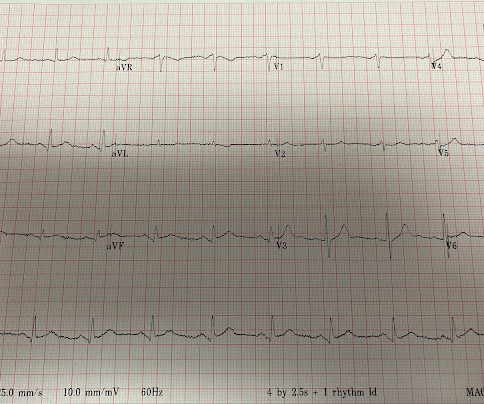
He denied any known medical history, specifically: coronary artery disease, hypertension, dyslipidemia, diabetes, heart failure, myocardial infarction, or any prior PCI/stent. Learning points 1] Acute Coronary Syndrome has many shades of clinical manifestation. No appreciable skin pallor. Here is the time-zero 12 Lead ECG.
During angiogram in the cath lab, the patient suffered two episodes of ventricular fibrillation for which he was successfully defibrillated. Angiogram showed a culprit lesion of 100% stenosis to the right coronary artery and 100% stenosis of the right posterior descending artery, both with TIMI 0 flow. Just another NSTEMI.
Here is the coronaryangiogram: A distal thrombotic right coronary artery (RCA) occlusion ! The lesion was successfully stented. Here is the post-intervention angiogram and post-PCI ECG. A significant amount of thrombotic material was aspirated by manual thrombectomy (see below for aspirated thrombi).
Diamond and Forrester accomplished this by first establishing the prevalence of coronary artery disease based on how clinically likely patients with chest pain symptoms were found to have coronary disease based on a coronaryangiogram.
Cardiology felt her chest pain to be, most likely, the result of coronary supply-demand mismatch in the context of HCM endothelial remodeling (i.e. Type II MI), however decided to pursue coronaryangiogram out of an abundance of caution. A mid-LAD culprit lesion was identified and stented.
Advanced multi-vessel disease was found with stents deployed to the mid-LCx (80% stenosis), D1 (90% stensosis), and the pLAD (95% stenosis). It’s judicious, then, to arrange for coronaryangiogram. Coronary occlusion, however, might be present concurrently with subendocardial ischemia on the time-zero ECG, or evolve into such.
The proximal and mid LAD stenoses were stented and the OM 2 was left alone. Heitner et al found that in 14% of patients with NSTEMI, a blinded interventional cardiologist interpreting coronaryangiograms identified a different culprit artery than CMR ( [link] ). Based on the EKGs, which lesion is most likely the culprit?
We organize all of the trending information in your field so you don't have to. Join thousands of users and stay up to date on the latest articles your peers are reading.
You know about us, now we want to get to know you!
Let's personalize your content
Let's get even more personalized
We recognize your account from another site in our network, please click 'Send Email' below to continue with verifying your account and setting a password.


Let's personalize your content