This site uses cookies to improve your experience. To help us insure we adhere to various privacy regulations, please select your country/region of residence. If you do not select a country, we will assume you are from the United States. Select your Cookie Settings or view our Privacy Policy and Terms of Use.
Cookie Settings
Cookies and similar technologies are used on this website for proper function of the website, for tracking performance analytics and for marketing purposes. We and some of our third-party providers may use cookie data for various purposes. Please review the cookie settings below and choose your preference.
Used for the proper function of the website
Used for monitoring website traffic and interactions
Cookie Settings
Cookies and similar technologies are used on this website for proper function of the website, for tracking performance analytics and for marketing purposes. We and some of our third-party providers may use cookie data for various purposes. Please review the cookie settings below and choose your preference.
Strictly Necessary: Used for the proper function of the website
Performance/Analytics: Used for monitoring website traffic and interactions
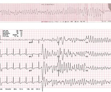
Workup including routine laboratory results, 12-lead electrocardiogram (ECG), echocardiogram, and coronaryangiogram was non-specific. During the intravenous lacosamide infusion, the patient developed sudden cardiac arrest caused by ventricular arrhythmias necessitating resuscitation.
Electrocardiogram (EKG) was unremarkable. A repeat coronaryangiogram was unremarkable. Circulation, Volume 150, Issue Suppl_1 , Page A4135360-A4135360, November 12, 2024. Case presentation:A 64-year-old man presented with one day of chest pain. Initial evaluation showed elevated cardiac enzymes (CE) and normal eosinophil count.
Explanation: Shown electrocardiogram suggests left ventricular hypertrophy. Shown electrocardiogram suggests left ventricular hypertrophy. Start aspirin and Plavix Correct answer: (B) (B) Echocardiogram is indicated. Hypertrophic cardiomyopathy is one of them. Start with a Free Trial.
She had a prior history of "NSTEMI" one month ago, during which she had a coronaryangiogram reportedly showing no stenosis in any coronary artery. Electromechanical association: a subtle electrocardiogram artifact. Her vitals were within normal limits. Acute chest pain and a bizarre ECG Bizarre (Hyperacute??)
Cardiology admitted him for observation with plans for next-day coronaryangiogram. Unfortunately, due to the patient’s abrupt exodus from the PCI center – without benefit of coronaryangiogram, or echo, for example – the disposition will forever remain unknown. [1] The peak Troponin I confirmed myocardial infarction. (A
Smith , d and Muzaffer Değertekin a DIFOCCULT: DIagnostic accuracy oF electrocardiogram for acute coronary OCClUsion resuLTing in myocardial infarction. His first electrocardiogram ( ECG) is given below: --Sinus bradycardia. Here is the coronaryangiogram: A distal thrombotic right coronary artery (RCA) occlusion !
Cardiology felt her chest pain to be, most likely, the result of coronary supply-demand mismatch in the context of HCM endothelial remodeling (i.e. Type II MI), however decided to pursue coronaryangiogram out of an abundance of caution. A mid-LAD culprit lesion was identified and stented. Pacing Clin Electrophysiol.
Incidence of an acute coronary occlusion. New insights into the use of the 12-lead electrocardiogram for diagnosing acute myocardial infarction in the emergency department. We investigated the incidence of an acutely occluded coronary in patients presenting with STE-aVR with multi-lead ST depression.
It’s judicious, then, to arrange for coronaryangiogram. Coronary occlusion, however, might be present concurrently with subendocardial ischemia on the time-zero ECG, or evolve into such. elevated BP), but rather directly correlated with coronary obstruction and stymied TIMI flow. Does the ECG normalize? 2] Aslanger, E.,
We organize all of the trending information in your field so you don't have to. Join thousands of users and stay up to date on the latest articles your peers are reading.
You know about us, now we want to get to know you!
Let's personalize your content
Let's get even more personalized
We recognize your account from another site in our network, please click 'Send Email' below to continue with verifying your account and setting a password.


Let's personalize your content