
6 Cardiology Board Review Questions That Will Help You Pass the Boards
BoardVitals - Cardiovascular
JUNE 14, 2017
Question banks are a favorite exam preparation resource for Cardiologists that want to practice in the format of the exam.

BoardVitals - Cardiovascular
JUNE 14, 2017
Question banks are a favorite exam preparation resource for Cardiologists that want to practice in the format of the exam.

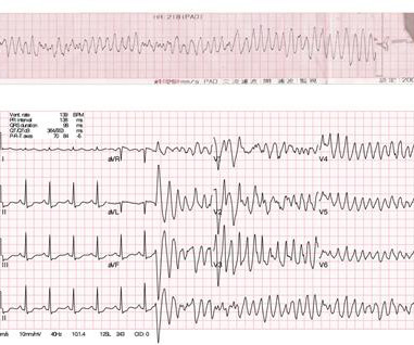
Circulation
NOVEMBER 11, 2024
Electrocardiogram (EKG) was unremarkable. A repeat coronary angiogram was unremarkable. Circulation, Volume 150, Issue Suppl_1 , Page A4135360-A4135360, November 12, 2024. Case presentation:A 64-year-old man presented with one day of chest pain. Initial evaluation showed elevated cardiac enzymes (CE) and normal eosinophil count.

Dr. Smith's ECG Blog
MARCH 17, 2023
She had a prior history of "NSTEMI" one month ago, during which she had a coronary angiogram reportedly showing no stenosis in any coronary artery. Electromechanical association: a subtle electrocardiogram artifact. Her vitals were within normal limits. Acute chest pain and a bizarre ECG Bizarre (Hyperacute??)

EMS 12-Lead
MAY 20, 2022
Cardiology felt her chest pain to be, most likely, the result of coronary supply-demand mismatch in the context of HCM endothelial remodeling (i.e. Type II MI), however decided to pursue coronary angiogram out of an abundance of caution. A mid-LAD culprit lesion was identified and stented. Pacing Clin Electrophysiol.

Dr. Smith's ECG Blog
NOVEMBER 4, 2022
Incidence of an acute coronary occlusion. New insights into the use of the 12-lead electrocardiogram for diagnosing acute myocardial infarction in the emergency department. We investigated the incidence of an acutely occluded coronary in patients presenting with STE-aVR with multi-lead ST depression.
Frontiers in Cardiovascular Medicine
JUNE 3, 2024
Workup including routine laboratory results, 12-lead electrocardiogram (ECG), echocardiogram, and coronary angiogram was non-specific. During the intravenous lacosamide infusion, the patient developed sudden cardiac arrest caused by ventricular arrhythmias necessitating resuscitation.

EMS 12-Lead
APRIL 8, 2022
Cardiology admitted him for observation with plans for next-day coronary angiogram. Unfortunately, due to the patient’s abrupt exodus from the PCI center – without benefit of coronary angiogram, or echo, for example – the disposition will forever remain unknown. [1] The peak Troponin I confirmed myocardial infarction. (A

Expert insights. Personalized for you.


Let's personalize your content